 Movie
Movie Controller

[English] 日本語
 Yorodumi
- PDB-3azu: X-RAY CRYSTAL STRUCTURE OF THE TWO SITE-SPECIFIC MUTANTS HIS35GLN... -
+ Open data

- Basic information
| Entry | Database: PDB / ID: 3azu | ||||||
|---|---|---|---|---|---|---|---|
| Title | X-RAY CRYSTAL STRUCTURE OF THE TWO SITE-SPECIFIC MUTANTS HIS35GLN AND HIS35LEU OF AZURIN FROM PSEUDOMONAS AERUGINOSA | ||||||
 Components Components | AZURIN | ||||||
 Keywords Keywords | ELECTRON TRANSFER(CUPROPROTEIN) | ||||||
| Function / homology |  Function and homology information Function and homology informationtransition metal ion binding / periplasmic space / electron transfer activity / copper ion binding / zinc ion binding / identical protein binding / plasma membrane Similarity search - Function | ||||||
| Biological species |   Pseudomonas aeruginosa (bacteria) Pseudomonas aeruginosa (bacteria) | ||||||
| Method |  X-RAY DIFFRACTION / Resolution: 2.1 Å X-RAY DIFFRACTION / Resolution: 2.1 Å | ||||||
 Authors Authors | Messerschmidt, A. / Nar, H. / Huber, R. | ||||||
 Citation Citation | Journal: J.Mol.Biol. / Year: 1991 Title: X-ray crystal structure of the two site-specific mutants His35Gln and His35Leu of azurin from Pseudomonas aeruginosa. Authors: Nar, H. / Messerschmidt, A. / Huber, R. / van de Kamp, M. / Canters, G.W. | ||||||
| History |
|
- Structure visualization
| Structure viewer | Molecule: MolmilJmol/JSmol |
|---|
- Downloads & links
-Download
| PDBx/mmCIF format | 3azu.cif.gz | 113.9 KB | Display | PDBx/mmCIF format |
|---|---|---|---|---|
| PDB format | pdb3azu.ent.gz | 88.5 KB | Display | PDB format |
| PDBx/mmJSON format | 3azu.json.gz | Tree view | PDBx/mmJSON format | |
| Others |  Other downloads Other downloads |
-Validation report
| Summary document | 3azu_validation.pdf.gz | 383.4 KB | Display | wwPDB validaton report |
|---|---|---|---|---|
| Full document | 3azu_full_validation.pdf.gz | 389.6 KB | Display | |
| Data in XML | 3azu_validation.xml.gz | 10.9 KB | Display | |
| Data in CIF | 3azu_validation.cif.gz | 18.6 KB | Display | |
| Arichive directory | https://data.pdbj.org/pub/pdb/validation_reports/az/3azuftp://data.pdbj.org/pub/pdb/validation_reports/az/3azu | HTTPS FTP |
-Related structure data

-Links
 PDBj
PDBj- Assembly
| Deposited unit | 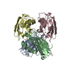
| ||||||||
|---|---|---|---|---|---|---|---|---|---|
| 1 |
| ||||||||
| Unit cell |
| ||||||||
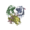
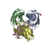
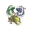
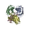
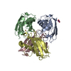
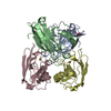
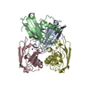
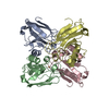
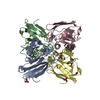
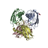
| Details | THERE ARE FOUR MOLECULES IN THE ASYMMETRIC UNIT WHICH ARE ARRANGED AS A DIMER OF DIMERS. |
-Components
| #1: Protein | Mass: 13950.796 Da / Num. of mol.: 4 / Mutation: H35Q Source method: isolated from a genetically manipulated source Source: (gene. exp.) Pseudomonas aeruginosa (bacteria) / References: UniProt: P00282#2: Chemical | ChemComp-CU /   Mass: 63.546 Da / Num. of mol.: 4 / Source method: obtained synthetically / Formula: Cu Mass: 63.546 Da / Num. of mol.: 4 / Source method: obtained synthetically / Formula: Cu#3: Water | ChemComp-HOH / |  Mass: 18.015 Da / Num. of mol.: 281 / Source method: isolated from a natural source / Formula: H2O Mass: 18.015 Da / Num. of mol.: 281 / Source method: isolated from a natural source / Formula: H2OHas protein modification | Y | |
|---|
-Experimental details
-Experiment
| Experiment | Method: X-RAY DIFFRACTION |
|---|
- Sample preparation
| Crystal | Density Matthews: 2.33 Å3/Da / Density % sol: 47.22 % | ||||||||||||||||||||||||||||||
|---|---|---|---|---|---|---|---|---|---|---|---|---|---|---|---|---|---|---|---|---|---|---|---|---|---|---|---|---|---|---|---|
| Crystal grow | *PLUS Method: vapor diffusion / PH range low: 5.7 / PH range high: 5.5 | ||||||||||||||||||||||||||||||
| Components of the solutions | *PLUS
|
-Data collection
| Radiation | Scattering type: x-ray |
|---|---|
| Radiation wavelength | Relative weight: 1 |
| Reflection | *PLUS Highest resolution: 2.09 Å / Lowest resolution: 9999 Å / Num. obs: 22537 / % possible obs: 66.6 % / Num. measured all: 99146 / Rmerge(I) obs: 0.097 |
| Reflection shell | *PLUS Highest resolution: 2.09 Å / Lowest resolution: 2.14 Å / % possible obs: 21.6 % |
- Processing
| Software |
| ||||||||||||
|---|---|---|---|---|---|---|---|---|---|---|---|---|---|
| Refinement | Rfactor Rwork: 0.163 / Highest resolution: 2.1 Å | ||||||||||||
| Refinement step | Cycle: LAST / Highest resolution: 2.1 Å
| ||||||||||||
| Refine LS restraints |
| ||||||||||||
| Refinement | *PLUS Highest resolution: 2.1 Å / Num. reflection obs: 20747 / Rfactor obs: 0.163 / Lowest resolution: 8 Å | ||||||||||||
| Solvent computation | *PLUS | ||||||||||||
| Displacement parameters | *PLUS | ||||||||||||
| Refine LS restraints | *PLUS
|
