 Movie
Movie Controller

[English] 日本語
 Yorodumi
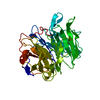
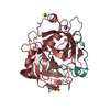
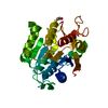
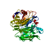
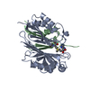
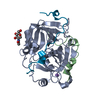
- PDB-3ams: Crystal Structures of Bacillus subtilis Alkaline Phytase in Compl... -
+ Open data

- Basic information
| Entry | Database: PDB / ID: 3ams | ||||||
|---|---|---|---|---|---|---|---|
| Title | Crystal Structures of Bacillus subtilis Alkaline Phytase in Complex with Ca2+, Cd2+, Co2+, Ni2+, Mg2+ and myo-Inositol Hexasulfate | ||||||
 Components Components | 3-phytase | ||||||
 Keywords Keywords | HYDROLASE/HYDROLASE INHIBITOR / beta-propeller / phytase / phytate / myo-Inositol hexasulfate / HYDROLASE-HYDROLASE INHIBITOR complex | ||||||
| Function / homology |  Function and homology information Function and homology information3-phytase / inositol hexakisphosphate 3-phosphatase activity / extracellular region Similarity search - Function | ||||||
| Biological species |  | ||||||
| Method |  X-RAY DIFFRACTION / SYNCHROTRON / MOLECULAR REPLACEMENT / Resolution: 2.08 Å X-RAY DIFFRACTION / SYNCHROTRON / MOLECULAR REPLACEMENT / Resolution: 2.08 Å | ||||||
 Authors Authors | Zeng, Y.F. / Ko, T.P. / Lai, H.L. / Cheng, Y.S. / Wu, T.H. / Ma, Y. / Yang, C.S. / Cheng, K.J. / Huang, C.H. / Guo, R.T. / Liu, J.R. | ||||||
 Citation Citation | Journal: J.Mol.Biol. / Year: 2011 Title: Crystal structures of Bacillus alkaline phytase in complex with divalent metal ions and inositol hexasulfate Authors: Zeng, Y.F. / Ko, T.P. / Lai, H.L. / Cheng, Y.S. / Wu, T.H. / Ma, Y. / Chen, C.C. / Yang, C.S. / Cheng, K.J. / Huang, C.H. / Guo, R.T. / Liu, J.R. | ||||||
| History |
|
- Structure visualization
| Structure viewer | Molecule: MolmilJmol/JSmol |
|---|
- Downloads & links
-Download
| PDBx/mmCIF format | 3ams.cif.gz | 91.3 KB | Display | PDBx/mmCIF format |
|---|---|---|---|---|
| PDB format | pdb3ams.ent.gz | 67 KB | Display | PDB format |
| PDBx/mmJSON format | 3ams.json.gz | Tree view | PDBx/mmJSON format | |
| Others |  Other downloads Other downloads |
-Validation report
| Summary document | 3ams_validation.pdf.gz | 1 MB | Display | wwPDB validaton report |
|---|---|---|---|---|
| Full document | 3ams_full_validation.pdf.gz | 1 MB | Display | |
| Data in XML | 3ams_validation.xml.gz | 19.2 KB | Display | |
| Data in CIF | 3ams_validation.cif.gz | 28.7 KB | Display | |
| Arichive directory | https://data.pdbj.org/pub/pdb/validation_reports/am/3amsftp://data.pdbj.org/pub/pdb/validation_reports/am/3ams | HTTPS FTP |
-Related structure data
| Related structure data |  3amrC 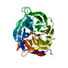 1h6lS C: citing same article ( S: Starting model for refinement |
|---|---|
| Similar structure data |


-Links
 PDBj
PDBj


- Assembly
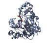
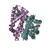
| Deposited unit | 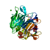
| ||||||||
|---|---|---|---|---|---|---|---|---|---|
| 1 |
| ||||||||
| Unit cell |
| ||||||||
| Components on special symmetry positions |
|
-Components
| #1: Protein | Mass: 39131.043 Da / Num. of mol.: 1 Source method: isolated from a genetically manipulated source Source: (gene. exp.) | ||||
|---|---|---|---|---|---|
| #2: Chemical | ChemComp-IHS / 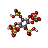  Mass: 660.535 Da / Num. of mol.: 1 / Source method: obtained synthetically / Formula: C6H12O24S6 Mass: 660.535 Da / Num. of mol.: 1 / Source method: obtained synthetically / Formula: C6H12O24S6 | ||||
| #3: Chemical | ChemComp-CA /   Mass: 40.078 Da / Num. of mol.: 4 / Source method: obtained synthetically / Formula: Ca Mass: 40.078 Da / Num. of mol.: 4 / Source method: obtained synthetically / Formula: Ca#4: Chemical | ChemComp-CD /   Mass: 112.411 Da / Num. of mol.: 7 / Source method: obtained synthetically / Formula: Cd Mass: 112.411 Da / Num. of mol.: 7 / Source method: obtained synthetically / Formula: Cd#5: Water | ChemComp-HOH / |  Mass: 18.015 Da / Num. of mol.: 335 / Source method: isolated from a natural source / Formula: H2O Mass: 18.015 Da / Num. of mol.: 335 / Source method: isolated from a natural source / Formula: H2O |
-Experimental details
-Experiment
| Experiment | Method: X-RAY DIFFRACTION / Number of used crystals: 1 |
|---|
- Sample preparation
| Crystal | Density Matthews: 2.22 Å3/Da / Density % sol: 44.57 % |
|---|---|
| Crystal grow | Temperature: 298 K / Method: vapor diffusion, sitting drop / pH: 7.5 Details: 5mM CoCl2, 5mM CdCl2, 5mM NiCl2, 5mM MgCl2, 0.1M HEPES, 12% PEG 3350 , pH 7.5, VAPOR DIFFUSION, SITTING DROP, temperature 298.0K |
-Data collection
| Diffraction | Mean temperature: 100 K |
|---|---|
| Diffraction source | Source: SYNCHROTRON / Site: NSRRC  / Beamline: BL13B1 / Wavelength: 1 Å / Beamline: BL13B1 / Wavelength: 1 Å |
| Detector | Type: ADSC QUANTUM 315 / Detector: CCD / Date: Dec 12, 2009 / Details: mirrors |
| Radiation | Monochromator: GRAPHITE / Protocol: SINGLE WAVELENGTH / Monochromatic (M) / Laue (L): M / Scattering type: x-ray |
| Radiation wavelength | Wavelength: 1 Å / Relative weight: 1 |
| Reflection | Resolution: 2.08→25 Å / Num. all: 21559 / Num. obs: 21329 / % possible obs: 99.6 % / Observed criterion σ(F): 0 / Observed criterion σ(I): 0 / Redundancy: 6.9 % / Rmerge(I) obs: 0.082 / Net I/σ(I): 26.2 |
| Reflection shell | Highest resolution: 2.08 Å / Redundancy: 6.6 % / Rmerge(I) obs: 0.281 / Mean I/σ(I) obs: 7.2 / Num. unique all: 82 / % possible all: 98.7 |
- Processing
| Software |
| ||||||||||||||||||||||||||||
|---|---|---|---|---|---|---|---|---|---|---|---|---|---|---|---|---|---|---|---|---|---|---|---|---|---|---|---|---|---|
| Refinement | Method to determine structure: MOLECULAR REPLACEMENT Starting model: PDB ENTRY 1H6L Resolution: 2.08→25 Å / Isotropic thermal model: Isotropic / Cross valid method: THROUGHOUT / σ(F): 0 / σ(I): 0 / Stereochemistry target values: Engh & Huber
| ||||||||||||||||||||||||||||
| Solvent computation | Bsol: 67.7355 Å2 | ||||||||||||||||||||||||||||
| Displacement parameters | Biso mean: 33.82 Å2
| ||||||||||||||||||||||||||||
| Refine analyze | Luzzati coordinate error obs: 0.19 Å / Luzzati sigma a obs: 0.17 Å | ||||||||||||||||||||||||||||
| Refinement step | Cycle: LAST / Resolution: 2.08→25 Å
| ||||||||||||||||||||||||||||
| Refine LS restraints |
| ||||||||||||||||||||||||||||
| LS refinement shell | Highest resolution: 2.08 Å
| ||||||||||||||||||||||||||||
| Xplor file |
|
