 Movie
Movie Controller

[English] 日本語
 Yorodumi
- PDB-5ohf: Globin sensor domain of AfGcHK (FeIII form) in complex with cyani... -
+ Open data

- Basic information
| Entry | Database: PDB / ID: 5ohf | ||||||||||||
|---|---|---|---|---|---|---|---|---|---|---|---|---|---|
| Title | Globin sensor domain of AfGcHK (FeIII form) in complex with cyanide, partially reduced | ||||||||||||
 Components Components | Globin-coupled histidine kinase | ||||||||||||
 Keywords Keywords | TRANSFERASE / Heme / Sensor protein / Oxygen sensor / Globin sensor domain / globin domain / cyanide | ||||||||||||
| Function / homology |  Function and homology information Function and homology informationpeptidyl-histidine phosphorylation / phosphorelay sensor kinase activity / histidine kinase / phosphorelay signal transduction system / oxygen binding / heme binding / protein homodimerization activity / ATP binding / metal ion binding Similarity search - Function | ||||||||||||
| Biological species | Anaeromyxobacter sp. | ||||||||||||
| Method |  X-RAY DIFFRACTION / SYNCHROTRON / MOLECULAR REPLACEMENT / Resolution: 1.8 Å X-RAY DIFFRACTION / SYNCHROTRON / MOLECULAR REPLACEMENT / Resolution: 1.8 Å | ||||||||||||
 Authors Authors | Skalova, T. / Kolenko, P. / Dohnalek, J. / Stranava, M. / Martinkova, M. | ||||||||||||
| Funding support |  Czech Republic, 3items Czech Republic, 3items
| ||||||||||||
 Citation Citation | Journal: J. Biol. Chem. / Year: 2017 Title: Coordination and redox state-dependent structural changes of the heme-based oxygen sensor AfGcHK associated with intraprotein signal transduction. Authors: Stranava, M. / Man, P. / Skalova, T. / Kolenko, P. / Blaha, J. / Fojtikova, V. / Martinek, V. / Dohnalek, J. / Lengalova, A. / Rosulek, M. / Shimizu, T. / Martinkova, M. | ||||||||||||
| History |
|
- Structure visualization
| Structure viewer | Molecule: MolmilJmol/JSmol |
|---|
- Downloads & links
-Download
| PDBx/mmCIF format | 5ohf.cif.gz | 306.2 KB | Display | PDBx/mmCIF format |
|---|---|---|---|---|
| PDB format | pdb5ohf.ent.gz | 252.1 KB | Display | PDB format |
| PDBx/mmJSON format | 5ohf.json.gz | Tree view | PDBx/mmJSON format | |
| Others |  Other downloads Other downloads |
-Validation report
| Arichive directory | https://data.pdbj.org/pub/pdb/validation_reports/oh/5ohfftp://data.pdbj.org/pub/pdb/validation_reports/oh/5ohf | HTTPS FTP |
|---|
-Related structure data
| Related structure data |  5oheSC S: Starting model for refinement C: citing same article ( |
|---|---|
| Similar structure data |


-Links
 PDBj
PDBj


- Assembly
| Deposited unit | 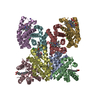
| ||||||||
|---|---|---|---|---|---|---|---|---|---|
| 1 | 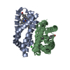
| ||||||||
| 2 | 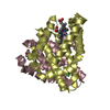
| ||||||||
| 3 | 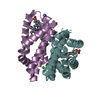
| ||||||||
| 4 | 
| ||||||||
| Unit cell |
|
-Components
-Protein , 1 types, 8 molecules ABCDEFGH
| #1: Protein | Mass: 18370.861 Da / Num. of mol.: 8 Source method: isolated from a genetically manipulated source Source: (gene. exp.)  Anaeromyxobacter sp. (strain Fw109-5) (bacteria) Anaeromyxobacter sp. (strain Fw109-5) (bacteria)Gene: gchK, Anae109_2438 / Plasmid: pET21c(+) / Production host: |
|---|
-Non-polymers , 5 types, 1038 molecules 


| #2: Chemical | ChemComp-HEM /  Mass: 616.487 Da / Num. of mol.: 8 / Source method: obtained synthetically / Formula: C34H32FeN4O4 Mass: 616.487 Da / Num. of mol.: 8 / Source method: obtained synthetically / Formula: C34H32FeN4O4#3: Chemical | ChemComp-CYN /  Mass: 26.017 Da / Num. of mol.: 8 / Source method: obtained synthetically / Formula: CN Mass: 26.017 Da / Num. of mol.: 8 / Source method: obtained synthetically / Formula: CN#4: Chemical | ChemComp-CL /  Mass: 35.453 Da / Num. of mol.: 5 / Source method: obtained synthetically / Formula: Cl Mass: 35.453 Da / Num. of mol.: 5 / Source method: obtained synthetically / Formula: Cl#5: Chemical | ChemComp-NA / |  Mass: 22.990 Da / Num. of mol.: 1 / Source method: obtained synthetically / Formula: Na Mass: 22.990 Da / Num. of mol.: 1 / Source method: obtained synthetically / Formula: Na#6: Water | ChemComp-HOH / | Mass: 18.015 Da / Num. of mol.: 1016 / Source method: isolated from a natural source / Formula: H2O |
|---|
-Experimental details
-Experiment
| Experiment | Method: X-RAY DIFFRACTION / Number of used crystals: 1 |
|---|
- Sample preparation
| Crystal | Density Matthews: 2.27 Å3/Da / Density % sol: 46 % Description: Crystal shape - red wedge block 300x80x80 um; the crystal was soaked for 15 min in a maternal drop containing additionally 10 mM sodium dithionite. |
|---|---|
| Crystal grow | Temperature: 298 K / Method: vapor diffusion, hanging drop / pH: 6.7 Details: 20.7% (w/v) PEG3350, 200 mM MgCl2, 0.01 M KCN, 7.5% (v/v) glycerol, 0.1 M MMT buffer system (DL-Malic acid, MES monohydrate, Tris, NaOH, HCl), pH 6.7 |
-Data collection
| Diffraction | Mean temperature: 100 K |
|---|---|
| Diffraction source | Source: SYNCHROTRON / Site: PETRA III, EMBL c/o DESY  / Beamline: P13 (MX1) / Wavelength: 1 Å / Beamline: P13 (MX1) / Wavelength: 1 Å |
| Detector | Type: DECTRIS PILATUS 6M / Detector: PIXEL / Date: Sep 11, 2015 |
| Radiation | Protocol: SINGLE WAVELENGTH / Monochromatic (M) / Laue (L): M / Scattering type: x-ray |
| Radiation wavelength | Wavelength: 1 Å / Relative weight: 1 |
| Reflection | Resolution: 1.8→49.17 Å / Num. obs: 127077 / % possible obs: 100 % / Observed criterion σ(I): -3.7 / Redundancy: 7.3 % / Biso Wilson estimate: 21 Å2 / CC1/2: 0.993 / Rmerge(I) obs: 0.059 / Rpim(I) all: 0.034 / Net I/σ(I): 16.7 |
| Reflection shell | Resolution: 1.8→1.83 Å / Redundancy: 7.6 % / Rmerge(I) obs: 0.761 / Mean I/σ(I) obs: 2.7 / Num. unique obs: 6192 / CC1/2: 0.844 / Rpim(I) all: 0.442 / % possible all: 100 |
- Processing
| Software |
| ||||||||||||||||||||||||||||||||||||||||||||||||||||||||||||||||||||||||||||||||||||||||||||||||||||||||||||||||||||||||||||||||||||||||||||||||||||||||||||||||||||||||||||||||||||||
|---|---|---|---|---|---|---|---|---|---|---|---|---|---|---|---|---|---|---|---|---|---|---|---|---|---|---|---|---|---|---|---|---|---|---|---|---|---|---|---|---|---|---|---|---|---|---|---|---|---|---|---|---|---|---|---|---|---|---|---|---|---|---|---|---|---|---|---|---|---|---|---|---|---|---|---|---|---|---|---|---|---|---|---|---|---|---|---|---|---|---|---|---|---|---|---|---|---|---|---|---|---|---|---|---|---|---|---|---|---|---|---|---|---|---|---|---|---|---|---|---|---|---|---|---|---|---|---|---|---|---|---|---|---|---|---|---|---|---|---|---|---|---|---|---|---|---|---|---|---|---|---|---|---|---|---|---|---|---|---|---|---|---|---|---|---|---|---|---|---|---|---|---|---|---|---|---|---|---|---|---|---|---|---|
| Refinement | Method to determine structure: MOLECULAR REPLACEMENT Starting model: 5OHE Resolution: 1.8→49.17 Å / Cor.coef. Fo:Fc: 0.958 / SU B: 2.748 / SU ML: 0.082 / Cross valid method: THROUGHOUT / ESU R: 0.138 / Details: HYDROGENS HAVE BEEN ADDED IN THE RIDING POSITIONS
| ||||||||||||||||||||||||||||||||||||||||||||||||||||||||||||||||||||||||||||||||||||||||||||||||||||||||||||||||||||||||||||||||||||||||||||||||||||||||||||||||||||||||||||||||||||||
| Solvent computation | Ion probe radii: 0.8 Å / Shrinkage radii: 0.8 Å / VDW probe radii: 1.2 Å | ||||||||||||||||||||||||||||||||||||||||||||||||||||||||||||||||||||||||||||||||||||||||||||||||||||||||||||||||||||||||||||||||||||||||||||||||||||||||||||||||||||||||||||||||||||||
| Displacement parameters | Biso mean: 28 Å2
| ||||||||||||||||||||||||||||||||||||||||||||||||||||||||||||||||||||||||||||||||||||||||||||||||||||||||||||||||||||||||||||||||||||||||||||||||||||||||||||||||||||||||||||||||||||||
| Refinement step | Cycle: 1 / Resolution: 1.8→49.17 Å
| ||||||||||||||||||||||||||||||||||||||||||||||||||||||||||||||||||||||||||||||||||||||||||||||||||||||||||||||||||||||||||||||||||||||||||||||||||||||||||||||||||||||||||||||||||||||
| Refine LS restraints |
|