 Movie
Movie Controller

[English] 日本語
 Yorodumi
- PDB-3am5: K316A mutant of Enoyl-ACP Reductase from Plasmodium falciparum (P... -
+ Open data

- Basic information
| Entry | Database: PDB / ID: 3am5 | ||||||
|---|---|---|---|---|---|---|---|
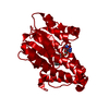
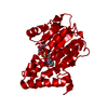
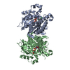
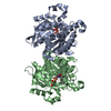
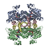
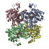
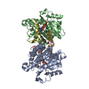
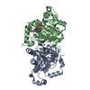
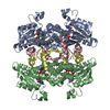
| Title | K316A mutant of Enoyl-ACP Reductase from Plasmodium falciparum (PfENR) in complex with triclosan | ||||||
 Components Components | Enoyl-ACP reductase | ||||||
 Keywords Keywords | OXIDOREDUCTASE/OXIDOREDUCTASE INHIBITOR / Triclosan / Enoyl-ACP reductase / mutant / oxidoreductase / P.falciparum / FabI / OXIDOREDUCTASE-OXIDOREDUCTASE INHIBITOR complex | ||||||
| Function / homology |  Function and homology information Function and homology informationenoyl-[acyl-carrier-protein] reductase (NADH) activity / fatty acid biosynthetic process / nucleotide binding Similarity search - Function | ||||||
| Biological species |  | ||||||
| Method |  X-RAY DIFFRACTION / MOLECULAR REPLACEMENT / Resolution: 2.05 Å X-RAY DIFFRACTION / MOLECULAR REPLACEMENT / Resolution: 2.05 Å | ||||||
 Authors Authors | Maity, K. / Banerjee, T. / Narayanappa, P. / Surolia, N. / Surolia, A. / Suguna, K. | ||||||
 Citation Citation | Journal: Iubmb Life / Year: 2011 Title: Effect of substrate binding loop mutations on the structure, kinetics, and inhibition of enoyl acyl carrier protein reductase from plasmodium falciparum Authors: Maity, K. / Banerjee, T. / Prabakaran, N. / Surolia, N. / Surolia, A. / Suguna, K. | ||||||
| History |
|
- Structure visualization
| Structure viewer | Molecule: MolmilJmol/JSmol |
|---|
- Downloads & links
-Download
| PDBx/mmCIF format | 3am5.cif.gz | 139.8 KB | Display | PDBx/mmCIF format |
|---|---|---|---|---|
| PDB format | pdb3am5.ent.gz | 107.8 KB | Display | PDB format |
| PDBx/mmJSON format | 3am5.json.gz | Tree view | PDBx/mmJSON format | |
| Others |  Other downloads Other downloads |
-Validation report
| Arichive directory | https://data.pdbj.org/pub/pdb/validation_reports/am/3am5ftp://data.pdbj.org/pub/pdb/validation_reports/am/3am5 | HTTPS FTP |
|---|
-Related structure data
| Related structure data |  3am3C  3am4C 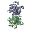 1uh5S S: Starting model for refinement C: citing same article ( |
|---|---|
| Similar structure data |


-Links
 PDBj
PDBj

- Assembly
| Deposited unit | 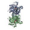
| ||||||||
|---|---|---|---|---|---|---|---|---|---|
| 1 | 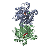
| ||||||||
| Unit cell |
|
-Components
| #1: Protein | Mass: 37186.086 Da / Num. of mol.: 2 / Fragment: residues 96-424 / Mutation: K316A Source method: isolated from a genetically manipulated source Source: (gene. exp.) Gene: fabI / Plasmid: PET-28a(+)(NOVAGEN) / Production host:  References: UniProt: Q9BJJ9, enoyl-[acyl-carrier-protein] reductase (NADH) #2: Chemical |   Mass: 663.425 Da / Num. of mol.: 2 / Source method: obtained synthetically / Formula: C21H27N7O14P2 / Comment: NAD*YM Mass: 663.425 Da / Num. of mol.: 2 / Source method: obtained synthetically / Formula: C21H27N7O14P2 / Comment: NAD*YM#3: Chemical |   Mass: 289.542 Da / Num. of mol.: 2 / Source method: obtained synthetically / Formula: C12H7Cl3O2 / Comment: antifungal, antibiotic, detergent*YM Mass: 289.542 Da / Num. of mol.: 2 / Source method: obtained synthetically / Formula: C12H7Cl3O2 / Comment: antifungal, antibiotic, detergent*YM#4: Water | ChemComp-HOH / |  Mass: 18.015 Da / Num. of mol.: 451 / Source method: isolated from a natural source / Formula: H2O Mass: 18.015 Da / Num. of mol.: 451 / Source method: isolated from a natural source / Formula: H2O |
|---|
-Experimental details
-Experiment
| Experiment | Method: X-RAY DIFFRACTION / Number of used crystals: 1 |
|---|
- Sample preparation
| Crystal | Density Matthews: 2.39 Å3/Da / Density % sol: 48.57 % |
|---|---|
| Crystal grow | Temperature: 289 K / Method: vapor diffusion, hanging drop / pH: 5.8 Details: 3M ammonium sulphate, 0.1M MES buffer, pH 5.8, VAPOR DIFFUSION, HANGING DROP, temperature 289K |
-Data collection
| Diffraction | Mean temperature: 100 K |
|---|---|
| Diffraction source | Source: ROTATING ANODE / Type: RIGAKU RU200 / Wavelength: 1.54 Å |
| Detector | Type: MAR scanner 345 mm plate / Detector: IMAGE PLATE / Date: Mar 18, 2009 / Details: Mirror |
| Radiation | Monochromator: OSMIC MIRROR / Protocol: SINGLE WAVELENGTH / Monochromatic (M) / Laue (L): M / Scattering type: x-ray |
| Radiation wavelength | Wavelength: 1.54 Å / Relative weight: 1 |
| Reflection | Resolution: 2.05→51.4 Å / Num. obs: 45800 / % possible obs: 100 % / Observed criterion σ(I): 0 / Redundancy: 9.8 % / Biso Wilson estimate: 26.6 Å2 / Rmerge(I) obs: 0.083 / Net I/σ(I): 20.3 |
| Reflection shell | Resolution: 2.05→2.16 Å / Redundancy: 9.7 % / Rmerge(I) obs: 0.461 / Mean I/σ(I) obs: 4.4 / Num. unique all: 6576 / % possible all: 100 |
- Processing
| Software |
| |||||||||||||||||||||||||||||||||||||||||||||||||||||||||||||||||
|---|---|---|---|---|---|---|---|---|---|---|---|---|---|---|---|---|---|---|---|---|---|---|---|---|---|---|---|---|---|---|---|---|---|---|---|---|---|---|---|---|---|---|---|---|---|---|---|---|---|---|---|---|---|---|---|---|---|---|---|---|---|---|---|---|---|---|
| Refinement | Method to determine structure: MOLECULAR REPLACEMENT Starting model: 1UH5 Resolution: 2.05→47.85 Å / Cor.coef. Fo:Fc: 0.964 / Cor.coef. Fo:Fc free: 0.943 / SU B: 3.554 / SU ML: 0.097 / Isotropic thermal model: RESTRAINED / Cross valid method: THROUGHOUT / σ(F): 0 / ESU R Free: 0.148 / Stereochemistry target values: MAXIMUM LIKELIHOOD / Details: BULK SOLVENT MODEL USED
| |||||||||||||||||||||||||||||||||||||||||||||||||||||||||||||||||
| Solvent computation | Ion probe radii: 0.8 Å / Shrinkage radii: 0.8 Å / VDW probe radii: 1.4 Å / Solvent model: BABINET MODEL WITH MASK | |||||||||||||||||||||||||||||||||||||||||||||||||||||||||||||||||
| Displacement parameters | Biso mean: 30.804 Å2
| |||||||||||||||||||||||||||||||||||||||||||||||||||||||||||||||||
| Refinement step | Cycle: LAST / Resolution: 2.05→47.85 Å
| |||||||||||||||||||||||||||||||||||||||||||||||||||||||||||||||||
| Refine LS restraints |
| |||||||||||||||||||||||||||||||||||||||||||||||||||||||||||||||||
| LS refinement shell | Resolution: 2.05→2.103 Å / Total num. of bins used: 20
|
