 Movie
Movie Controller

[English] 日本語
 Yorodumi
- PDB-1nhg: CRYSTAL STRUCTURE ANALYSIS OF PLASMODIUM FALCIPARUM ENOYL-ACYL-CA... -
+ Open data

- Basic information
| Entry | Database: PDB / ID: 1nhg | ||||||
|---|---|---|---|---|---|---|---|
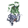
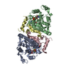
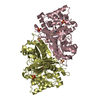
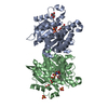
| Title | CRYSTAL STRUCTURE ANALYSIS OF PLASMODIUM FALCIPARUM ENOYL-ACYL-CARRIER-PROTEIN REDUCTASE WITH TRICLOSAN | ||||||
 Components Components | (enoyl-acyl carrier reductase) x 2 | ||||||
 Keywords Keywords | OXIDOREDUCTASE / Rossmann fold / short chain dehydrogenase reductase / nadh | ||||||
| Function / homology |  Function and homology information Function and homology informationenoyl-[acyl-carrier-protein] reductase (NADH) activity / fatty acid biosynthetic process / nucleotide binding Similarity search - Function | ||||||
| Biological species |  | ||||||
| Method |  X-RAY DIFFRACTION / MOLECULAR REPLACEMENT / Resolution: 2.43 Å X-RAY DIFFRACTION / MOLECULAR REPLACEMENT / Resolution: 2.43 Å | ||||||
 Authors Authors | Perozzo, R. / Kuo, M. / Sidhu, A.S. / Valiyaveettil, J.T. / Bittman, R. / Jacobs Jr., W.R. / Fidock, D.A. / Sacchettini, J.C. | ||||||
 Citation Citation | Journal: J.Biol.Chem. / Year: 2002 Title: Structural Elucidation of the Specificity of the Antibacterial Agent Triclosan for Malarial Enoyl Acyl Carrier Protein Reductase Authors: Perozzo, R. / Kuo, M. / Sidhu, A.S. / Valiyaveettil, J.T. / Bittman, R. / Jacobs Jr., W.R. / Fidock, D.A. / Sacchettini, J.C. | ||||||
| History |
|
- Structure visualization
| Structure viewer | Molecule: MolmilJmol/JSmol |
|---|
- Downloads & links
-Download
| PDBx/mmCIF format | 1nhg.cif.gz | 125.5 KB | Display | PDBx/mmCIF format |
|---|---|---|---|---|
| PDB format | pdb1nhg.ent.gz | 99.6 KB | Display | PDB format |
| PDBx/mmJSON format | 1nhg.json.gz | Tree view | PDBx/mmJSON format | |
| Others |  Other downloads Other downloads |
-Validation report
| Arichive directory | https://data.pdbj.org/pub/pdb/validation_reports/nh/1nhgftp://data.pdbj.org/pub/pdb/validation_reports/nh/1nhg | HTTPS FTP |
|---|
-Related structure data


-Links
 PDBj
PDBj

- Assembly
| Deposited unit | 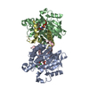
| ||||||||
|---|---|---|---|---|---|---|---|---|---|
| 1 |
| ||||||||
| 2 | 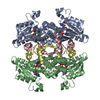
| ||||||||
| Unit cell |
|
-Components
| #1: Protein | Mass: 25744.570 Da / Num. of mol.: 2 Source method: isolated from a genetically manipulated source Source: (gene. exp.) Description: PET28A; / Plasmid: Plasmid / Production host: Bacteria (eubacteria) / Variant (production host): BL21(DE3) CodonPlus-RIL References: UniProt: Q9BH77, enoyl-[acyl-carrier-protein] reductase (NADH) #2: Protein | Mass: 6829.676 Da / Num. of mol.: 2 Source method: isolated from a genetically manipulated source Source: (gene. exp.) Description: pET28a / Plasmid: Plasmid / Production host: Bacteria (eubacteria) / Variant (production host): BL21(DE3) CodonPlus-RIL References: UniProt: Q9BH77, enoyl-[acyl-carrier-protein] reductase (NADH) #3: Chemical |   Mass: 663.425 Da / Num. of mol.: 2 / Source method: obtained synthetically / Formula: C21H27N7O14P2 / Comment: NAD*YM Mass: 663.425 Da / Num. of mol.: 2 / Source method: obtained synthetically / Formula: C21H27N7O14P2 / Comment: NAD*YM#4: Chemical |   Mass: 289.542 Da / Num. of mol.: 2 / Source method: obtained synthetically / Formula: C12H7Cl3O2 / Comment: antifungal, antibiotic, detergent*YM Mass: 289.542 Da / Num. of mol.: 2 / Source method: obtained synthetically / Formula: C12H7Cl3O2 / Comment: antifungal, antibiotic, detergent*YM |
|---|
-Experimental details
-Experiment
| Experiment | Method: X-RAY DIFFRACTION / Number of used crystals: 1 |
|---|
- Sample preparation
| Crystal | Density Matthews: 2.86 Å3/Da / Density % sol: 56.98 % | ||||||||||||||||||||||||||||||||||||||||||||||||||||||||
|---|---|---|---|---|---|---|---|---|---|---|---|---|---|---|---|---|---|---|---|---|---|---|---|---|---|---|---|---|---|---|---|---|---|---|---|---|---|---|---|---|---|---|---|---|---|---|---|---|---|---|---|---|---|---|---|---|---|
| Crystal grow | Temperature: 282 K / Method: vapor diffusion, hanging drop / pH: 5.6 Details: 2.35 M (NH4)2SO4, 100 mM sodium acetate pH 5.6, VAPOR DIFFUSION, HANGING DROP, temperature 282K | ||||||||||||||||||||||||||||||||||||||||||||||||||||||||
| Crystal grow | *PLUS pH: 7.5 | ||||||||||||||||||||||||||||||||||||||||||||||||||||||||
| Components of the solutions | *PLUS
|
-Data collection
| Diffraction | Mean temperature: 120 K |
|---|---|
| Diffraction source | Source: ROTATING ANODE / Type: RIGAKU RU200 / Wavelength: 1.5418 |
| Detector | Type: MACSCIENCE / Detector: IMAGE PLATE / Date: Nov 17, 2000 |
| Radiation | Protocol: SINGLE WAVELENGTH / Monochromatic (M) / Laue (L): M / Scattering type: x-ray |
| Radiation wavelength | Wavelength: 1.5418 Å / Relative weight: 1 |
| Reflection | Resolution: 2.43→30 Å / Num. all: 28104 / Num. obs: 28104 / % possible obs: 96.9 % / Observed criterion σ(F): 2 / Observed criterion σ(I): 0 |
| Reflection | *PLUS Num. obs: 29010 / Redundancy: 5.8 % / Rmerge(I) obs: 0.095 |
- Processing
| Software |
| ||||||||||||||||||||
|---|---|---|---|---|---|---|---|---|---|---|---|---|---|---|---|---|---|---|---|---|---|
| Refinement | Method to determine structure: MOLECULAR REPLACEMENT / Resolution: 2.43→30 Å / Cross valid method: THROUGHOUT / σ(F): 2 / σ(I): 0 / Stereochemistry target values: Engh & Huber
| ||||||||||||||||||||
| Refinement step | Cycle: LAST / Resolution: 2.43→30 Å
| ||||||||||||||||||||
| Refinement | *PLUS Lowest resolution: 30 Å / % reflection Rfree: 10 % | ||||||||||||||||||||
| Solvent computation | *PLUS | ||||||||||||||||||||
| Displacement parameters | *PLUS |
