 Movie
Movie Controller


+ Open data

- Basic information
| Entry | Database: PDB / ID: 1v35 | ||||||
|---|---|---|---|---|---|---|---|
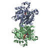
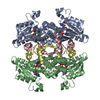
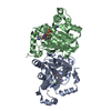
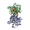
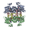
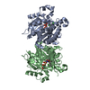
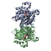
| Title | Crystal Structure of Eoyl-ACP Reductase with NADH | ||||||
 Components Components | enoyl-ACP reductase | ||||||
 Keywords Keywords | OXIDOREDUCTASE / FabI / NADH / ENOYL-ACP Reductase / P.falciparum | ||||||
| Function / homology |  Function and homology information Function and homology informationenoyl-[acyl-carrier-protein] reductase (NADH) activity / fatty acid biosynthetic process / nucleotide binding Similarity search - Function | ||||||
| Biological species |  | ||||||
| Method |  X-RAY DIFFRACTION / MOLECULAR REPLACEMENT / Resolution: 2.5 Å X-RAY DIFFRACTION / MOLECULAR REPLACEMENT / Resolution: 2.5 Å | ||||||
 Authors Authors | SwarnaMukhi, P.L. / Kapoor, M. / surolia, N. / Surolia, A. / Suguna, K. | ||||||
 Citation Citation | Journal: J.Mol.Biol. / Year: 2004 Title: Structural basis for the variation in triclosan affinity to enoyl reductases. Authors: Pidugu, L.S. / Kapoor, M. / Surolia, N. / Surolia, A. / Suguna, K. #1: Journal: Biochem.J. / Year: 2004 Title: Kinetic and structural analysis of the increased affinity of enoyl-ACP (acyl-carrier protein) reductase for triclosan in the presence of NAD+ Authors: Kapoor, M. / Swarnamukhi, P.L. / Surolia, N. / Suguna, K. / Surolia, A. | ||||||
| History |
| ||||||
| Remark 750 | TURN Determination method: author determined |
- Structure visualization
| Structure viewer | Molecule: MolmilJmol/JSmol |
|---|
- Downloads & links
-Download
| PDBx/mmCIF format | 1v35.cif.gz | 129.1 KB | Display | PDBx/mmCIF format |
|---|---|---|---|---|
| PDB format | pdb1v35.ent.gz | 99.4 KB | Display | PDB format |
| PDBx/mmJSON format | 1v35.json.gz | Tree view | PDBx/mmJSON format | |
| Others |  Other downloads Other downloads |
-Validation report
| Arichive directory | https://data.pdbj.org/pub/pdb/validation_reports/v3/1v35ftp://data.pdbj.org/pub/pdb/validation_reports/v3/1v35 | HTTPS FTP |
|---|
-Related structure data
| Related structure data |  1uh5C 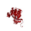 1enoS C: citing same article ( S: Starting model for refinement |
|---|---|
| Similar structure data |


-Links
 PDBj
PDBj


- Assembly
| Deposited unit | 
| ||||||||
|---|---|---|---|---|---|---|---|---|---|
| 1 | 
| ||||||||
| Unit cell |
| ||||||||
| Details | the other two subunits of the functional tetramer are generated by a-y,b-x,c/2-z |
-Components
| #1: Protein | Mass: 37244.184 Da / Num. of mol.: 2 / Fragment: residues 96-424 Source method: isolated from a genetically manipulated source Source: (gene. exp.) Plasmid: pET28A(+) / Species (production host): Escherichia coli / Production host:  References: UniProt: Q6LFB9, UniProt: Q9BJJ9*PLUS, enoyl-[acyl-carrier-protein] reductase (NADH) #2: Chemical |   Mass: 665.441 Da / Num. of mol.: 2 / Source method: obtained synthetically / Formula: C21H29N7O14P2 Mass: 665.441 Da / Num. of mol.: 2 / Source method: obtained synthetically / Formula: C21H29N7O14P2#3: Water | ChemComp-HOH / |  Mass: 18.015 Da / Num. of mol.: 178 / Source method: isolated from a natural source / Formula: H2O Mass: 18.015 Da / Num. of mol.: 178 / Source method: isolated from a natural source / Formula: H2O |
|---|
-Experimental details
-Experiment
| Experiment | Method: X-RAY DIFFRACTION / Number of used crystals: 1 |
|---|
- Sample preparation
| Crystal | Density Matthews: 2.88 Å3/Da / Density % sol: 57.02 % |
|---|---|
| Crystal grow | Temperature: 298 K / Method: vapor diffusion, hanging drop / pH: 5.6 Details: MES, Ammonium Sulphate, pH 5.6, VAPOR DIFFUSION, HANGING DROP, temperature 298.0K |
-Data collection
| Diffraction | Mean temperature: 298 K |
|---|---|
| Diffraction source | Source: ROTATING ANODE / Type: RIGAKU / Wavelength: 1.5418 Å |
| Detector | Type: RIGAKU / Detector: IMAGE PLATE / Details: osmic mirror |
| Radiation | Monochromator: osmic mirrors / Protocol: SINGLE WAVELENGTH / Monochromatic (M) / Laue (L): M / Scattering type: x-ray |
| Radiation wavelength | Wavelength: 1.5418 Å / Relative weight: 1 |
| Reflection | Resolution: 2.5→20 Å / Num. all: 26750 / Num. obs: 26266 / % possible obs: 98.2 % / Observed criterion σ(F): 0 / Observed criterion σ(I): 0 / Biso Wilson estimate: 24.3 Å2 / Rmerge(I) obs: 0.101 / Net I/σ(I): 14 |
| Reflection shell | Resolution: 2.5→2.59 Å / Rmerge(I) obs: 0.39 / Mean I/σ(I) obs: 5.7 / Num. unique all: 2527 / % possible all: 99.3 |
- Processing
| Software |
| ||||||||||||||||||||||||||||||||||||
|---|---|---|---|---|---|---|---|---|---|---|---|---|---|---|---|---|---|---|---|---|---|---|---|---|---|---|---|---|---|---|---|---|---|---|---|---|---|
| Refinement | Method to determine structure: MOLECULAR REPLACEMENT Starting model: 1ENO Resolution: 2.5→19.87 Å / Rfactor Rfree error: 0.005 / Data cutoff high absF: 336140.39 / Data cutoff low absF: 0 / Isotropic thermal model: RESTRAINED / Cross valid method: THROUGHOUT / σ(F): 0 / σ(I): 0 / Stereochemistry target values: Engh & Huber
| ||||||||||||||||||||||||||||||||||||
| Solvent computation | Solvent model: FLAT MODEL / Bsol: 59.9036 Å2 / ksol: 0.362163 e/Å3 | ||||||||||||||||||||||||||||||||||||
| Displacement parameters | Biso mean: 33.6 Å2
| ||||||||||||||||||||||||||||||||||||
| Refine analyze |
| ||||||||||||||||||||||||||||||||||||
| Refinement step | Cycle: LAST / Resolution: 2.5→19.87 Å
| ||||||||||||||||||||||||||||||||||||
| Refine LS restraints |
| ||||||||||||||||||||||||||||||||||||
| LS refinement shell | Resolution: 2.5→2.66 Å / Rfactor Rfree error: 0.018 / Total num. of bins used: 6
| ||||||||||||||||||||||||||||||||||||
| Xplor file |
|
