 Movie
Movie Controller

[English] 日本語
 Yorodumi
- PDB-3ahu: Crystal structure of YmaH (Hfq) from Bacillus subtilis in complex... -
+ Open data

- Basic information
| Entry | Database: PDB / ID: 3ahu | ||||||
|---|---|---|---|---|---|---|---|
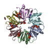
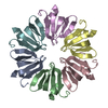
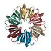
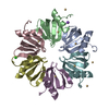
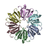
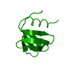
| Title | Crystal structure of YmaH (Hfq) from Bacillus subtilis in complex with an RNA aptamer. | ||||||
 Components Components |
| ||||||
 Keywords Keywords | TRANSLATION/RNA / Sm-like motif / Protein-RNA complex / TRANSLATION-RNA complex | ||||||
| Function / homology |  Function and homology information Function and homology informationregulation of translation, ncRNA-mediated / regulation of RNA stability / regulation of DNA-templated transcription / RNA binding / cytosol Similarity search - Function | ||||||
| Biological species |  | ||||||
| Method |  X-RAY DIFFRACTION / SYNCHROTRON / MOLECULAR REPLACEMENT / Resolution: 2.2 Å X-RAY DIFFRACTION / SYNCHROTRON / MOLECULAR REPLACEMENT / Resolution: 2.2 Å | ||||||
 Authors Authors | Baba, S. / Someya, T. / Kumasaka, T. / Kawai, G. / Nakamura, K. | ||||||
 Citation Citation | Journal: To be Published Title: Crystal structure of YmaH (Hfq) from Bacillus subtilis in complex with an RNA aptamer. Authors: Someya, T. / Baba, S. / Fujimoto, M. / Kumasaka, T. / Kawai, G. / Nakamura, K. #1: Journal: Acta Crystallogr.,Sect.F / Year: 2010 Title: Expression, crystallization and preliminary crystallographic analysis of RNA-binding protein Hfq (YmaH) from Bacillus subtilis in complex with an RNA aptamer. Authors: Baba, S. / Someya, T. / Kawai, G. / Nakamura, K. / Kumasaka, T. | ||||||
| History |
|
- Structure visualization
| Structure viewer | Molecule: MolmilJmol/JSmol |
|---|
- Downloads & links
-Download
| PDBx/mmCIF format | 3ahu.cif.gz | 56.9 KB | Display | PDBx/mmCIF format |
|---|---|---|---|---|
| PDB format | pdb3ahu.ent.gz | 40.9 KB | Display | PDB format |
| PDBx/mmJSON format | 3ahu.json.gz | Tree view | PDBx/mmJSON format | |
| Others |  Other downloads Other downloads |
-Validation report
| Summary document | 3ahu_validation.pdf.gz | 468.8 KB | Display | wwPDB validaton report |
|---|---|---|---|---|
| Full document | 3ahu_full_validation.pdf.gz | 482.9 KB | Display | |
| Data in XML | 3ahu_validation.xml.gz | 12 KB | Display | |
| Data in CIF | 3ahu_validation.cif.gz | 15.6 KB | Display | |
| Arichive directory | https://data.pdbj.org/pub/pdb/validation_reports/ah/3ahuftp://data.pdbj.org/pub/pdb/validation_reports/ah/3ahu | HTTPS FTP |
-Related structure data
| Related structure data |  1kq2S S: Starting model for refinement |
|---|---|
| Similar structure data |


-Links
 PDBj
PDBj

- Assembly
| Deposited unit | 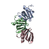
| ||||||||
|---|---|---|---|---|---|---|---|---|---|
| 1 | 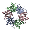
| ||||||||
| Unit cell |
|
-Components
| #1: Protein | Mass: 8934.214 Da / Num. of mol.: 3 Source method: isolated from a genetically manipulated source Source: (gene. exp.) #2: RNA chain | | Mass: 1978.277 Da / Num. of mol.: 1 / Source method: obtained synthetically #3: Water | ChemComp-HOH / |  Mass: 18.015 Da / Num. of mol.: 25 / Source method: isolated from a natural source / Formula: H2O Mass: 18.015 Da / Num. of mol.: 25 / Source method: isolated from a natural source / Formula: H2O |
|---|
-Experimental details
-Experiment
| Experiment | Method: X-RAY DIFFRACTION / Number of used crystals: 1 |
|---|
- Sample preparation
| Crystal | Density Matthews: 2.12 Å3/Da / Density % sol: 42.03 % |
|---|---|
| Crystal grow | Temperature: 293 K / Method: vapor diffusion, hanging drop / pH: 6.5 Details: 0.01M cobalt (II) chloride, 0.2M MES, 1.8M ammonium sulfate, pH 6.5, VAPOR DIFFUSION, HANGING DROP, temperature 293K |
-Data collection
| Diffraction | Mean temperature: 100 K |
|---|---|
| Diffraction source | Source: SYNCHROTRON / Site: SPring-8  / Beamline: BL38B1 / Wavelength: 1 Å / Beamline: BL38B1 / Wavelength: 1 Å |
| Detector | Type: RIGAKU JUPITER 210 / Detector: CCD / Date: Oct 17, 2008 / Details: Toroidal Mirror |
| Radiation | Monochromator: Fixed exit Si 111 double crystal monochromator Protocol: SINGLE WAVELENGTH / Monochromatic (M) / Laue (L): M / Scattering type: x-ray |
| Radiation wavelength | Wavelength: 1 Å / Relative weight: 1 |
| Reflection | Resolution: 2.2→30 Å / Num. all: 12626 / Num. obs: 12626 / % possible obs: 100 % / Observed criterion σ(F): 0 / Observed criterion σ(I): -3 / Redundancy: 7.4 % / Rmerge(I) obs: 0.033 / Rsym value: 0.029 / Net I/σ(I): 45.75 |
| Reflection shell | Resolution: 2.2→2.28 Å / Redundancy: 7.4 % / Rmerge(I) obs: 0.307 / Mean I/σ(I) obs: 4.84 / Num. unique all: 1219 / Rsym value: 0.248 / % possible all: 100 |
- Processing
| Software |
| |||||||||||||||||||||||||
|---|---|---|---|---|---|---|---|---|---|---|---|---|---|---|---|---|---|---|---|---|---|---|---|---|---|---|
| Refinement | Method to determine structure: MOLECULAR REPLACEMENT Starting model: 1KQ2 Resolution: 2.2→30 Å / Isotropic thermal model: RESTRAINED / Cross valid method: THROUGHOUT / σ(F): 0 / Stereochemistry target values: MAXIMUM LIKELIHOOD
| |||||||||||||||||||||||||
| Refinement step | Cycle: LAST / Resolution: 2.2→30 Å
|
