 Movie
Movie Controller

[English] 日本語
 Yorodumi
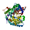
- PDB-3afq: Crystal structure of the single-stranded DNA binding protein from... -
+ Open data

- Basic information
| Entry | Database: PDB / ID: 3afq | ||||||
|---|---|---|---|---|---|---|---|
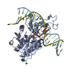
| Title | Crystal structure of the single-stranded DNA binding protein from Mycobacterium leprae (Form II) | ||||||
 Components Components | Single-stranded DNA-binding protein | ||||||
 Keywords Keywords | DNA BINDING PROTEIN / OB-fold / quaternary structure and stability / changes on oligomerisation / water-bridges / DNA damage / DNA repair / DNA replication / DNA-binding | ||||||
| Function / homology |  Function and homology information Function and homology information | ||||||
| Biological species |  Mycobacterium leprae (bacteria) Mycobacterium leprae (bacteria) | ||||||
| Method |  X-RAY DIFFRACTION / MOLECULAR REPLACEMENT / Resolution: 2.8 Å X-RAY DIFFRACTION / MOLECULAR REPLACEMENT / Resolution: 2.8 Å | ||||||
 Authors Authors | Kaushal, P.S. / Singh, P. / Sharma, A. / Muniyappa, K. / Vijayan, M. | ||||||
 Citation Citation | Journal: Acta Crystallogr.,Sect.D / Year: 2010 Title: X-ray and molecular-dynamics studies on Mycobacterium leprae single-stranded DNA-binding protein and comparison with other eubacterial SSB structures Authors: Kaushal, P.S. / Singh, P. / Sharma, A. / Muniyappa, K. / Vijayan, M. #1: Journal: Acta Crystallogr.,Sect.D / Year: 2005Title: Structure of Mycobacterium smegmatis single-stranded DNA-binding protein and a comparative study involving homologus SSBs: biological implications of structural plasticity and variability in quaternary association Authors: Saikrishnan, K. / Manjunath, G.P. / Singh, P. / Jeyakanthan, J. / Dauter, Z. / Sekar, K. / Muniyappa, K. / Vijayan, M. #2: Journal: J.Mol.Biol. / Year: 2003Title: Structure of Mycobacterium tuberculosis single-stranded DNA-binding protein. Variability in quaternary structure and its implications Authors: Saikrishnan, K. / Jeyakanthan, J. / Venkatesh, J. / Acharya, N. / Sekar, K. / Varshney, U. / Vijayan, M. #3: Journal: Nat.Struct.Biol. / Year: 1997Title: Crystal structure of human mitochondrial single-stranded DNA binding protein at 2.4 A resolution Authors: Yang, C. / Curth, U. / Urbanke, C. / Kang, C. #4: Journal: Proc.Natl.Acad.Sci.USA / Year: 1997Title: Crystal structure of the homo-tetrameric DNA binding domain of Escherichia coli single-stranded DNA-binding protein determined by multiwavelength x-ray diffraction on the selenomethionyl ...Title: Crystal structure of the homo-tetrameric DNA binding domain of Escherichia coli single-stranded DNA-binding protein determined by multiwavelength x-ray diffraction on the selenomethionyl protein at 2.9-A resolution Authors: Raghunathan, S. / Ricard, C.S. / Lohman, T.M. / Waksman, G. | ||||||
| History |
|
- Structure visualization
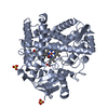
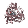
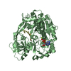
| Structure viewer | Molecule: MolmilJmol/JSmol |
|---|
- Downloads & links
-Download
| PDBx/mmCIF format | 3afq.cif.gz | 97.7 KB | Display | PDBx/mmCIF format |
|---|---|---|---|---|
| PDB format | pdb3afq.ent.gz | 73.6 KB | Display | PDB format |
| PDBx/mmJSON format | 3afq.json.gz | Tree view | PDBx/mmJSON format | |
| Others |  Other downloads Other downloads |
-Validation report
| Arichive directory | https://data.pdbj.org/pub/pdb/validation_reports/af/3afqftp://data.pdbj.org/pub/pdb/validation_reports/af/3afq | HTTPS FTP |
|---|
-Related structure data
| Related structure data |  3afpSC S: Starting model for refinement C: citing same article ( |
|---|---|
| Similar structure data |


-Links
 PDBj
PDBj- Assembly
| Deposited unit | 
| |||||||||||||||||||||||||||||||||||||||||||||||||||||||||||||||||||||||||||||||||||||||||||||||||||||||||||||||||||||||||||||||||||||||||||||||||||||||||
|---|---|---|---|---|---|---|---|---|---|---|---|---|---|---|---|---|---|---|---|---|---|---|---|---|---|---|---|---|---|---|---|---|---|---|---|---|---|---|---|---|---|---|---|---|---|---|---|---|---|---|---|---|---|---|---|---|---|---|---|---|---|---|---|---|---|---|---|---|---|---|---|---|---|---|---|---|---|---|---|---|---|---|---|---|---|---|---|---|---|---|---|---|---|---|---|---|---|---|---|---|---|---|---|---|---|---|---|---|---|---|---|---|---|---|---|---|---|---|---|---|---|---|---|---|---|---|---|---|---|---|---|---|---|---|---|---|---|---|---|---|---|---|---|---|---|---|---|---|---|---|---|---|---|---|
| 1 |
| |||||||||||||||||||||||||||||||||||||||||||||||||||||||||||||||||||||||||||||||||||||||||||||||||||||||||||||||||||||||||||||||||||||||||||||||||||||||||
| Unit cell |
| |||||||||||||||||||||||||||||||||||||||||||||||||||||||||||||||||||||||||||||||||||||||||||||||||||||||||||||||||||||||||||||||||||||||||||||||||||||||||
| Noncrystallographic symmetry (NCS) | NCS domain:
NCS domain segments: Ens-ID: 1 / Refine code: 2
|