- PDB-3afp: Crystal structure of the single-stranded DNA binding protein from... -
+
データを開く
IDまたはキーワード:
読み込み中...
-
基本情報
登録情報
データベース: PDB / ID: 3afp
タイトル
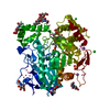
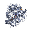
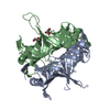
Crystal structure of the single-stranded DNA binding protein from Mycobacterium leprae (Form I)
要素
Single-stranded DNA-binding protein
キーワード
DNA BINDING PROTEIN / OB-fold / quaternary structure and stability / changes on oligomerisation / water-bridges / DNA damage / DNA repair / DNA replication / DNA-binding
機能・相同性
機能・相同性情報
nucleoid / single-stranded DNA binding / DNA replication 類似検索 - 分子機能
#1: ジャーナル: Acta Crystallogr.,Sect.D / 年: 2005 タイトル: Structure of Mycobacterium smegmatis single-stranded DNA-binding protein and a comparative study involving homologus SSBs: biological implications of structural plasticity and ...タイトル: Structure of Mycobacterium smegmatis single-stranded DNA-binding protein and a comparative study involving homologus SSBs: biological implications of structural plasticity and variability in quaternary association 著者: Saikrishnan, K. / Manjunath, G.P. / Singh, P. / Jeyakanthan, J. / Dauter, Z. / Sekar, K. / Muniyappa, K. / Vijayan, M.
#4: ジャーナル: Proc.Natl.Acad.Sci.USA / 年: 1997 タイトル: Crystal structure of the homo-tetrameric DNA binding domain of Escherichia coli single-stranded DNA-binding protein determined by multiwavelength x-ray diffraction on the ...タイトル: Crystal structure of the homo-tetrameric DNA binding domain of Escherichia coli single-stranded DNA-binding protein determined by multiwavelength x-ray diffraction on the selenomethionyl protein at 2.9-A resolution 著者: Raghunathan, S. / Ricard, C.S. / Lohman, T.M. / Waksman, G.
 ムービー
ムービー コントローラー


 データを開く
 基本情報
基本情報 要素
要素 キーワード
キーワード 機能・相同性情報
 Mycobacterium leprae (らい菌)
 X線回折 /
  データ登録者
 引用
引用 構造の表示
構造の表示 ダウンロードとリンク
 その他のダウンロード


 PDBj
PDBj 集合体
集合体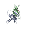

 分子量: 92.094 Da / 分子数: 1 / 由来タイプ: 合成 / 式: C3H8O3
 分子量: 112.411 Da / 分子数: 1 / 由来タイプ: 合成 / 式: Cd
 分子量: 18.015 Da / 分子数: 217 / 由来タイプ: 天然 / 式: H2O
 試料調製
試料調製 解析
解析