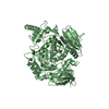
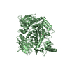
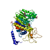
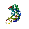
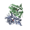
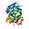
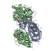
- PDB-3a9e: Crystal structure of a mixed agonist-bound RAR-alpha and antagoni... -
+
Open data
ID or keywords:
Loading...
-
Basic information
Entry
Database: PDB / ID: 3a9e
Title
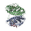
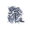
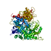
Crystal structure of a mixed agonist-bound RAR-alpha and antagonist-bound RXR-alpha heterodimer ligand binding domains
Components
(Retinoic acid receptor ...) x 2
13-mer (LXXLL motif) from Nuclear receptor coactivator 2
Keywords
TRANSCRIPTION / Nucleus / Receptor / Transcription regulation / Structural Genomics / SPINE2-complexes / Structural Proteomics in Europe
Function / homology
Function and homology information
Transcriptional regulation of granulopoiesis / Carnitine shuttle / Transcriptional regulation of white adipocyte differentiation / Signaling by Retinoic Acid / Sertoli cell fate commitment / positive regulation of binding / NR1H2 & NR1H3 regulate gene expression to control bile acid homeostasis / NR1H3 & NR1H2 regulate gene expression linked to cholesterol transport and efflux / SUMOylation of intracellular receptors / trachea cartilage development ...Transcriptional regulation of granulopoiesis / Carnitine shuttle / Transcriptional regulation of white adipocyte differentiation / Signaling by Retinoic Acid / Sertoli cell fate commitment / positive regulation of binding / NR1H2 & NR1H3 regulate gene expression to control bile acid homeostasis / NR1H3 & NR1H2 regulate gene expression linked to cholesterol transport and efflux / SUMOylation of intracellular receptors / trachea cartilage development / Recycling of bile acids and salts / Synthesis of bile acids and bile salts / glandular epithelial cell development / visceral serous pericardium development / Nuclear Receptor transcription pathway / Synthesis of bile acids and bile salts via 7alpha-hydroxycholesterol / Synthesis of bile acids and bile salts via 27-hydroxycholesterol / ventricular cardiac muscle cell differentiation / mesenchyme development / chondroblast differentiation / Endogenous sterols / embryonic camera-type eye development / positive regulation of translational initiation by iron / maternal placenta development / MLL4 and MLL3 complexes regulate expression of PPARG target genes in adipogenesis and hepatic steatosis / growth plate cartilage development / protein kinase B binding / positive regulation of T-helper 2 cell differentiation / prostate gland development / angiogenesis involved in coronary vascular morphogenesis / negative regulation of granulocyte differentiation / Regulation of lipid metabolism by PPARalpha / retinoic acid-responsive element binding / negative regulation of cartilage development / Cytoprotection by HMOX1 / regulation of hematopoietic progenitor cell differentiation / nuclear protein quality control by the ubiquitin-proteasome system / secretory columnal luminar epithelial cell differentiation involved in prostate glandular acinus development / positive regulation of thyroid hormone receptor signaling pathway / positive regulation of interleukin-5 production / positive regulation of interleukin-13 production / camera-type eye development / retinoic acid binding / nuclear retinoic acid receptor binding / outflow tract septum morphogenesis / cardiac muscle cell differentiation / positive regulation of vitamin D receptor signaling pathway / TGFBR3 expression / response to vitamin A / nuclear vitamin D receptor binding / apoptotic cell clearance / Signaling by Retinoic Acid / nuclear thyroid hormone receptor binding / heterocyclic compound binding / ureteric bud development / RNA polymerase II intronic transcription regulatory region sequence-specific DNA binding / regulation of branching involved in prostate gland morphogenesis / regulation of myelination / DNA-binding transcription repressor activity / DNA binding domain binding / ventricular cardiac muscle tissue morphogenesis / limb development / LBD domain binding / locomotor rhythm / positive regulation of interleukin-4 production / positive regulation of lipoprotein transport / face development / nuclear steroid receptor activity / aryl hydrocarbon receptor binding / protein kinase A binding / negative regulation of type II interferon production / germ cell development / cellular response to Thyroglobulin triiodothyronine / regulation of glucose metabolic process / Synthesis of bile acids and bile salts / regulation of lipid metabolic process / alpha-actinin binding / cellular response to estrogen stimulus / negative regulation of tumor necrosis factor production / Synthesis of bile acids and bile salts via 27-hydroxycholesterol / Endogenous sterols / Synthesis of bile acids and bile salts via 7alpha-hydroxycholesterol / monocyte differentiation / response to retinoic acid / cellular response to low-density lipoprotein particle stimulus / positive regulation of bone mineralization / cardiac muscle cell proliferation / Recycling of bile acids and salts / transcription regulator inhibitor activity / retinoic acid receptor signaling pathway / heart morphogenesis / cellular response to hormone stimulus / cellular response to retinoic acid / Regulation of lipid metabolism by PPARalpha / cell maturation / peptide binding / peroxisome proliferator activated receptor signaling pathway / response to progesterone / hormone-mediated signaling pathway / positive regulation of cell cycle Similarity search - Function
In the structure databanks used in Yorodumi, some data are registered as the other names, "COVID-19 virus" and "2019-nCoV". Here are the details of the virus and the list of structure data.
Jan 31, 2019. EMDB accession codes are about to change! (news from PDBe EMDB page)
EMDB accession codes are about to change! (news from PDBe EMDB page)
The allocation of 4 digits for EMDB accession codes will soon come to an end. Whilst these codes will remain in use, new EMDB accession codes will include an additional digit and will expand incrementally as the available range of codes is exhausted. The current 4-digit format prefixed with “EMD-” (i.e. EMD-XXXX) will advance to a 5-digit format (i.e. EMD-XXXXX), and so on. It is currently estimated that the 4-digit codes will be depleted around Spring 2019, at which point the 5-digit format will come into force.
The EM Navigator/Yorodumi systems omit the EMD- prefix.
Related info.:Q: What is EMD? / ID/Accession-code notation in Yorodumi/EM Navigator
Yorodumi is a browser for structure data from EMDB, PDB, SASBDB, etc.
This page is also the successor to EM Navigator detail page, and also detail information page/front-end page for Omokage search.
The word "yorodu" (or yorozu) is an old Japanese word meaning "ten thousand". "mi" (miru) is to see.
Related info.:EMDB / PDB / SASBDB / Comparison of 3 databanks / Yorodumi Search / Aug 31, 2016. New EM Navigator & Yorodumi / Yorodumi Papers / Jmol/JSmol / Function and homology information / Changes in new EM Navigator and Yorodumi
 Movie
Movie Controller

 Yorodumi
 Open data
 Basic information
 Components
 Keywords
 Function and homology information
 Homo sapiens (human)
 X-RAY DIFFRACTION /
  Authors
 Citation
 Structure visualization
 Downloads & links
 Other downloads
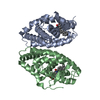


 PDBj
PDBj


 Assembly
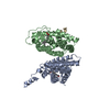

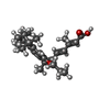

 Mass: 396.562 Da / Num. of mol.: 1 / Source method: obtained synthetically / Formula: C26H36O3
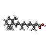
 Mass: 300.435 Da / Num. of mol.: 1 / Source method: obtained synthetically / Formula: C20H28O2
 Sample preparation
 / Beamline: ID23-1 / Wavelength: 1.0723 Å
 Processing