 Movie
Movie Controller

[English] 日本語
 Yorodumi
- PDB-1b0z: The crystal structure of phosphoglucose isomerase-an enzyme with ... -
+ Open data

- Basic information
| Entry | Database: PDB / ID: 1b0z | ||||||
|---|---|---|---|---|---|---|---|
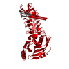
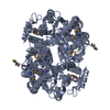
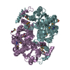
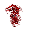
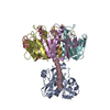
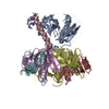
| Title | The crystal structure of phosphoglucose isomerase-an enzyme with autocrine motility factor activity in tumor cells | ||||||
 Components Components | PROTEIN (PHOSPHOGLUCOSE ISOMERASE) | ||||||
 Keywords Keywords | ISOMERASE / PHOSPHOGLUCOSE ISOMERASE / AUTOCRINEFACTOR / NEUROLEUKIN / CRYSTALLOGRAPHY MOTILITY | ||||||
| Function / homology |  Function and homology information Function and homology informationglucose-6-phosphate isomerase / glucose-6-phosphate isomerase activity / glucose 6-phosphate metabolic process / carbohydrate derivative binding / monosaccharide binding / gluconeogenesis / glycolytic process / cytosol Similarity search - Function | ||||||
| Biological species |   Geobacillus stearothermophilus (bacteria) Geobacillus stearothermophilus (bacteria) | ||||||
| Method |  X-RAY DIFFRACTION / MIRAS / Resolution: 2.3 Å X-RAY DIFFRACTION / MIRAS / Resolution: 2.3 Å | ||||||
 Authors Authors | Sun, Y.-J. / Chou, C.-C. / Chen, W.-S. / Meng, M. / Hsiao, C.-D. | ||||||
 Citation Citation | Journal: J.Biol.Chem. / Year: 2000 Title: The crystal structure of phosphoglucose isomerase/autocrine motility factor/neuroleukin complexed with its carbohydrate phosphate inhibitors suggests its substrate/receptor recognition Authors: Chou, C.-C. / Sun, Y.-J. / Meng, M. / Hsiao, C.-D. #1: Journal: J.Mol.Biol. / Year: 1977Title: Crystallographic Structure Analysis of Glucose-6-Phosphate Isomerase at 3.5 A Resolution Authors: Shaw, P.J. / Muirhead, H. | ||||||
| History |
|
- Structure visualization
| Structure viewer | Molecule: MolmilJmol/JSmol |
|---|
- Downloads & links
-Download
| PDBx/mmCIF format | 1b0z.cif.gz | 101 KB | Display | PDBx/mmCIF format |
|---|---|---|---|---|
| PDB format | pdb1b0z.ent.gz | 78.6 KB | Display | PDB format |
| PDBx/mmJSON format | 1b0z.json.gz | Tree view | PDBx/mmJSON format | |
| Others |  Other downloads Other downloads |
-Validation report
| Arichive directory | https://data.pdbj.org/pub/pdb/validation_reports/b0/1b0zftp://data.pdbj.org/pub/pdb/validation_reports/b0/1b0z | HTTPS FTP |
|---|
-Related structure data


-Links
 PDBj
PDBj

- Assembly
| Deposited unit | 
| ||||||||
|---|---|---|---|---|---|---|---|---|---|
| 1 | 
| ||||||||
| 2 | 
| ||||||||
| Unit cell |
|
-Components
| #1: Protein | Mass: 50202.758 Da / Num. of mol.: 1 Source method: isolated from a genetically manipulated source Source: (gene. exp.) Geobacillus stearothermophilus (bacteria)Gene: PGIB / Plasmid: PMMB67EH / Production host: |
|---|---|
| #2: Water | ChemComp-HOH /  Mass: 18.015 Da / Num. of mol.: 184 / Source method: isolated from a natural source / Formula: H2O Mass: 18.015 Da / Num. of mol.: 184 / Source method: isolated from a natural source / Formula: H2O |
-Experimental details
-Experiment
| Experiment | Method: X-RAY DIFFRACTION / Number of used crystals: 1 |
|---|
- Sample preparation
| Crystal | Density Matthews: 3.08 Å3/Da / Density % sol: 60 % |
|---|---|
| Crystal grow | Method: vapor diffusion, hanging drop / pH: 7 Details: CRYSTALS WERE GROWN BY HANGING DROP VAPOR DIFFUSION FROM 4-UL DROPLETS OF PROTEIN SOLUTION (15 MG/ML) IN 50 MM PHOSPHATE BUFFER (PH7.0) AND 0.2 M AMMONIUM PHOSPHATE AGAINST A RESERVOIR OF ...Details: CRYSTALS WERE GROWN BY HANGING DROP VAPOR DIFFUSION FROM 4-UL DROPLETS OF PROTEIN SOLUTION (15 MG/ML) IN 50 MM PHOSPHATE BUFFER (PH7.0) AND 0.2 M AMMONIUM PHOSPHATE AGAINST A RESERVOIR OF THE ABOVE BUFFER CONTAINING 0.4M AMMONIUM PHOSPHATE., VAPOR DIFFUSION, HANGING DROP |
| Crystal grow | *PLUS Method: unknown |
-Data collection
| Diffraction | Mean temperature: 287 K |
|---|---|
| Diffraction source | Source: ROTATING ANODE / Type: RIGAKU RU300 / Wavelength: 1.5418 |
| Detector | Type: RIGAKU RAXIS II / Detector: IMAGE PLATE / Date: Jul 25, 1997 / Details: MIRRORS |
| Radiation | Protocol: SINGLE WAVELENGTH / Monochromatic (M) / Laue (L): M / Scattering type: x-ray |
| Radiation wavelength | Wavelength: 1.5418 Å / Relative weight: 1 |
| Reflection | Resolution: 2.3→30.7 Å / Num. obs: 24024 / % possible obs: 93.9 % / Observed criterion σ(I): 2 / Redundancy: 4.3 % / Biso Wilson estimate: 21.11 Å2 / Rmerge(I) obs: 0.07 / Net I/σ(I): 12 |
| Reflection shell | Resolution: 2.3→2.38 Å / Redundancy: 3.1 % / Rmerge(I) obs: 0.36 / Mean I/σ(I) obs: 3.2 / % possible all: 83.4 |
- Processing
| Software |
| ||||||||||||||||||||||||||||||||||||||||||||||||||||||||||||
|---|---|---|---|---|---|---|---|---|---|---|---|---|---|---|---|---|---|---|---|---|---|---|---|---|---|---|---|---|---|---|---|---|---|---|---|---|---|---|---|---|---|---|---|---|---|---|---|---|---|---|---|---|---|---|---|---|---|---|---|---|---|
| Refinement | Method to determine structure: MIRAS / Resolution: 2.3→8 Å / Rfactor Rfree error: 0.013 / Data cutoff high absF: 1000000 / Data cutoff low absF: 0.001 / Cross valid method: THROUGHOUT / σ(F): 2
| ||||||||||||||||||||||||||||||||||||||||||||||||||||||||||||
| Displacement parameters | Biso mean: 21.62 Å2 | ||||||||||||||||||||||||||||||||||||||||||||||||||||||||||||
| Refine analyze | Luzzati coordinate error obs: 0.23 Å / Luzzati d res low obs: 8 Å / Luzzati sigma a obs: 0.19 Å | ||||||||||||||||||||||||||||||||||||||||||||||||||||||||||||
| Refinement step | Cycle: LAST / Resolution: 2.3→8 Å
| ||||||||||||||||||||||||||||||||||||||||||||||||||||||||||||
| Refine LS restraints |
| ||||||||||||||||||||||||||||||||||||||||||||||||||||||||||||
| LS refinement shell | Resolution: 2.3→2.4 Å / Rfactor Rfree error: 0.035 / Total num. of bins used: 8
| ||||||||||||||||||||||||||||||||||||||||||||||||||||||||||||
| Xplor file | Serial no: 1 / Param file: PARHCSDX.PRO / Topol file: TOPHCSDX.PRO |
