 Movie
Movie Controller

[English] 日本語
 Yorodumi
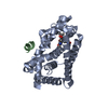
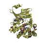
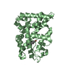
- PDB-3a01: Crystal structure of Aristaless and Clawless homeodomains bound to DNA -
+ Open data

- Basic information
| Entry | Database: PDB / ID: 3a01 | ||||||
|---|---|---|---|---|---|---|---|
| Title | Crystal structure of Aristaless and Clawless homeodomains bound to DNA | ||||||
 Components Components |
| ||||||
 Keywords Keywords | Gene Regulation/DNA / HOMEODOMAIN / PROTEIN-DNA COMPLEX / DNA-binding / Homeobox / Nucleus / Developmental protein / Gene Regulation-DNA COMPLEX | ||||||
| Function / homology |  Function and homology information Function and homology informationelongation of arista core / leg disc development / antennal morphogenesis / imaginal disc-derived leg morphogenesis / chaeta development / neuron development / RNA polymerase II transcription regulatory region sequence-specific DNA binding / DNA-binding transcription factor activity, RNA polymerase II-specific / negative regulation of DNA-templated transcription / regulation of transcription by RNA polymerase II ...elongation of arista core / leg disc development / antennal morphogenesis / imaginal disc-derived leg morphogenesis / chaeta development / neuron development / RNA polymerase II transcription regulatory region sequence-specific DNA binding / DNA-binding transcription factor activity, RNA polymerase II-specific / negative regulation of DNA-templated transcription / regulation of transcription by RNA polymerase II / positive regulation of DNA-templated transcription / protein-containing complex / DNA binding / nucleus Similarity search - Function | ||||||
| Biological species |  | ||||||
| Method |  X-RAY DIFFRACTION / SYNCHROTRON / MOLECULAR REPLACEMENT / Resolution: 2.7 Å X-RAY DIFFRACTION / SYNCHROTRON / MOLECULAR REPLACEMENT / Resolution: 2.7 Å | ||||||
 Authors Authors | Miyazono, K. / Nagata, K. / Saigo, K. / Kojima, T. / Tanokura, M. | ||||||
 Citation Citation | Journal: Embo J. / Year: 2010 Title: Cooperative DNA-binding and sequence-recognition mechanism of aristaless and clawless Authors: Miyazono, K. / Zhi, Y. / Takamura, Y. / Nagata, K. / Saigo, K. / Kojima, T. / Tanokura, M. | ||||||
| History |
|
- Structure visualization
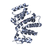
| Structure viewer | Molecule: MolmilJmol/JSmol |
|---|
- Downloads & links
-Download
| PDBx/mmCIF format | 3a01.cif.gz | 107 KB | Display | PDBx/mmCIF format |
|---|---|---|---|---|
| PDB format | pdb3a01.ent.gz | 78.6 KB | Display | PDB format |
| PDBx/mmJSON format | 3a01.json.gz | Tree view | PDBx/mmJSON format | |
| Others |  Other downloads Other downloads |
-Validation report
| Arichive directory | https://data.pdbj.org/pub/pdb/validation_reports/a0/3a01ftp://data.pdbj.org/pub/pdb/validation_reports/a0/3a01 | HTTPS FTP |
|---|
-Related structure data
| Related structure data |  3a02SC  3a03SC 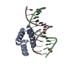 3lnqC  1pufS S: Starting model for refinement C: citing same article ( |
|---|---|
| Similar structure data |


-Links
 PDBj
PDBj


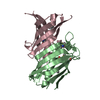
- Assembly
| Deposited unit | 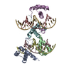
| ||||||||
|---|---|---|---|---|---|---|---|---|---|
| 1 | 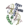
| ||||||||
| 2 | 
| ||||||||
| Unit cell |
|
-Components
| #1: Protein | Mass: 11191.974 Da / Num. of mol.: 2 / Fragment: Clawless Homeobox, residues 170-261 Source method: isolated from a genetically manipulated source Source: (gene. exp.)  #2: Protein | Mass: 8335.609 Da / Num. of mol.: 2 / Fragment: Homeobox, residues 80-146 Source method: isolated from a genetically manipulated source Source: (gene. exp.) #3: DNA chain | Mass: 5257.419 Da / Num. of mol.: 2 / Source method: obtained synthetically #4: DNA chain | Mass: 5155.375 Da / Num. of mol.: 2 / Source method: obtained synthetically |
|---|
-Experimental details
-Experiment
| Experiment | Method: X-RAY DIFFRACTION / Number of used crystals: 1 |
|---|
- Sample preparation
| Crystal | Density Matthews: 2.8 Å3/Da / Density % sol: 56.06 % |
|---|
-Data collection
| Diffraction | Mean temperature: 100 K |
|---|---|
| Diffraction source | Source: SYNCHROTRON / Site: Photon Factory  / Beamline: AR-NW12A / Wavelength: 1 Å / Beamline: AR-NW12A / Wavelength: 1 Å |
| Detector | Type: ADSC QUANTUM 210 / Detector: CCD |
| Radiation | Protocol: SINGLE WAVELENGTH / Monochromatic (M) / Laue (L): M / Scattering type: x-ray |
| Radiation wavelength | Wavelength: 1 Å / Relative weight: 1 |
| Reflection | Resolution: 2.7→20 Å / Num. obs: 18995 |
- Processing
| Software |
| ||||||||||||||||||||||||||||||||||||||||||||||||||||||||||||||||||||||||||||||||||||||||||||||||||||||||||||||||||||||||||||||||||||||||||||||||||||||||||||||||||||||||||
|---|---|---|---|---|---|---|---|---|---|---|---|---|---|---|---|---|---|---|---|---|---|---|---|---|---|---|---|---|---|---|---|---|---|---|---|---|---|---|---|---|---|---|---|---|---|---|---|---|---|---|---|---|---|---|---|---|---|---|---|---|---|---|---|---|---|---|---|---|---|---|---|---|---|---|---|---|---|---|---|---|---|---|---|---|---|---|---|---|---|---|---|---|---|---|---|---|---|---|---|---|---|---|---|---|---|---|---|---|---|---|---|---|---|---|---|---|---|---|---|---|---|---|---|---|---|---|---|---|---|---|---|---|---|---|---|---|---|---|---|---|---|---|---|---|---|---|---|---|---|---|---|---|---|---|---|---|---|---|---|---|---|---|---|---|---|---|---|---|---|---|---|
| Refinement | Method to determine structure: MOLECULAR REPLACEMENT Starting model: PDB ENTRIES 3A02, 3A03, and 1PUF Resolution: 2.7→19.65 Å / Cor.coef. Fo:Fc: 0.936 / Cor.coef. Fo:Fc free: 0.904 / SU ML: 0.279 / Cross valid method: THROUGHOUT / ESU R: 0.793 / ESU R Free: 0.367 / Stereochemistry target values: MAXIMUM LIKELIHOOD
| ||||||||||||||||||||||||||||||||||||||||||||||||||||||||||||||||||||||||||||||||||||||||||||||||||||||||||||||||||||||||||||||||||||||||||||||||||||||||||||||||||||||||||
| Solvent computation | Ion probe radii: 0.8 Å / Shrinkage radii: 0.8 Å / VDW probe radii: 1.2 Å / Solvent model: MASK | ||||||||||||||||||||||||||||||||||||||||||||||||||||||||||||||||||||||||||||||||||||||||||||||||||||||||||||||||||||||||||||||||||||||||||||||||||||||||||||||||||||||||||
| Refinement step | Cycle: LAST / Resolution: 2.7→19.65 Å
| ||||||||||||||||||||||||||||||||||||||||||||||||||||||||||||||||||||||||||||||||||||||||||||||||||||||||||||||||||||||||||||||||||||||||||||||||||||||||||||||||||||||||||
| Refine LS restraints |
| ||||||||||||||||||||||||||||||||||||||||||||||||||||||||||||||||||||||||||||||||||||||||||||||||||||||||||||||||||||||||||||||||||||||||||||||||||||||||||||||||||||||||||
| LS refinement shell | Resolution: 2.7→2.769 Å / Total num. of bins used: 20
|
