[日本語] English
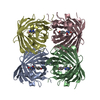
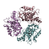
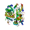
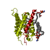
- PDB-2zz9: Structure of aquaporin-4 S180D mutant at 2.8 A resolution by elec... -

+
データを開く


IDまたはキーワード:

読み込み中...

-
基本情報

登録情報
データベース: PDB / ID: 2zz9
タイトルStructure of aquaporin-4 S180D mutant at 2.8 A resolution by electron crystallography
要素Aquaporin-4
キーワードTRANSPORT PROTEIN / WATER TRANSPORT / WATER CHANNEL / AQUAPORIN / TWO-DIMENSIONAL CRYSTAL / MEMBRANE PROTEIN / BACULOVIRUS EXPRESSION SYSTEM / Glycoprotein / Membrane / Phosphoprotein / Transmembrane / Transport
機能・相同性
機能・相同性情報


Passive transport by Aquaporins / cerebrospinal fluid secretion / renal water absorption / regulation of vascular endothelial growth factor production / cerebrospinal fluid circulation / astrocyte end-foot / intracellular water homeostasis / water transport / water channel activity / negative regulation of cell adhesion molecule production ...Passive transport by Aquaporins / cerebrospinal fluid secretion / renal water absorption / regulation of vascular endothelial growth factor production / cerebrospinal fluid circulation / astrocyte end-foot / intracellular water homeostasis / water transport / water channel activity / negative regulation of cell adhesion molecule production / cell projection membrane / multicellular organismal-level water homeostasis / Vasopressin regulates renal water homeostasis via Aquaporins / cellular response to interleukin-6 / negative regulation of interleukin-1 beta production / negative regulation of interleukin-6 production / cellular response to interleukin-1 / response to glucocorticoid / T-tubule / basal plasma membrane / cellular response to estradiol stimulus / establishment of localization in cell / female pregnancy / cellular response to glucose stimulus / sensory perception of sound / carbon dioxide transport / sarcolemma / cellular response to type II interferon / cell-cell adhesion / cell-cell junction / basolateral plasma membrane / protein homotetramerization / endosome membrane / external side of plasma membrane / protein-containing complex / extracellular region / identical protein binding / plasma membrane / cytoplasm
類似検索 - 分子機能
Glycerol uptake facilitator protein / Glycerol uptake facilitator protein. / Aquaporin transporter / Major intrinsic protein, conserved site / MIP family signature. / Major intrinsic protein / Major intrinsic protein / Aquaporin-like / Up-down Bundle / Mainly Alpha
類似検索 - ドメイン・相同性
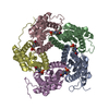
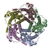
1,2-dioleoyl-sn-glycero-3-phosphoethanolamine / Aquaporin-4
類似検索 - 構成要素
生物種Rattus norvegicus (ドブネズミ)
手法電子線結晶学 / 分子置換 / クライオ電子顕微鏡法 / 解像度: 2.8 Å
データ登録者Tani, K. / Mitsuma, T. / Hiroaki, Y. / Kamegawa, A. / Nishikawa, K. / Tanimura, Y. / Fujiyoshi, Y.
引用ジャーナル: J Mol Biol / : 2009
タイトル: Mechanism of aquaporin-4's fast and highly selective water conduction and proton exclusion.
著者: Kazutoshi Tani / Tadanori Mitsuma / Yoko Hiroaki / Akiko Kamegawa / Kouki Nishikawa / Yukihiro Tanimura / Yoshinori Fujiyoshi /
要旨: Members of the aquaporin (AQP) family are expressed in almost every organism, including 13 homologues in humans. Based on the electron crystallographic structure of AQP1, the hydrogen-bond isolation ...Members of the aquaporin (AQP) family are expressed in almost every organism, including 13 homologues in humans. Based on the electron crystallographic structure of AQP1, the hydrogen-bond isolation mechanism was proposed to explain why AQPs are impermeable to protons despite their very fast water conduction. The mechanism by which AQPs exclude protons remained controversial, however. Here we present the structure of AQP4 at 2.8 A resolution obtained by electron crystallography of double-layered two-dimensional crystals. The resolution has been improved from the previous 3.2 A, with accompanying improvement in data quality resulting in the ability to identify individual water molecules. Our structure of AQP4, the predominant water channel in the brain, reveals eight water molecules in the channel. The arrangement of the waters provides support for the hydrogen-bond isolation mechanism. Our AQP4 structure also visualizes five lipids, showing that direct interactions of the extracellular surface of AQP4 with three lipids in the adjoining membrane help stabilize the membrane junction.
履歴
登録2009年2月6日登録サイト: PDBJ / 処理サイト: PDBJ
改定 1.02009年6月9日Provider: repository / タイプ: Initial release
改定 1.12011年7月13日Group: Version format compliance
改定 1.22014年10月29日Group: Non-polymer description
改定 2.02021年11月10日Group: Advisory / Atomic model ...Advisory / Atomic model / Author supporting evidence / Data collection / Database references / Derived calculations / Experimental preparation / Non-polymer description / Structure summary
カテゴリ: atom_site / atom_site_anisotrop ...atom_site / atom_site_anisotrop / chem_comp / database_2 / diffrn_radiation_wavelength / diffrn_source / em_image_scans / em_single_particle_entity / em_vitrification / entity / pdbx_entity_nonpoly / pdbx_unobs_or_zero_occ_atoms / struct_ref_seq_dif / struct_site
Item: _atom_site.B_iso_or_equiv / _atom_site.Cartn_x ..._atom_site.B_iso_or_equiv / _atom_site.Cartn_x / _atom_site.Cartn_y / _atom_site.Cartn_z / _atom_site.auth_atom_id / _atom_site.label_atom_id / _atom_site.type_symbol / _atom_site_anisotrop.U[1][1] / _atom_site_anisotrop.U[1][2] / _atom_site_anisotrop.U[1][3] / _atom_site_anisotrop.U[2][2] / _atom_site_anisotrop.U[2][3] / _atom_site_anisotrop.U[3][3] / _atom_site_anisotrop.pdbx_auth_atom_id / _atom_site_anisotrop.pdbx_label_atom_id / _atom_site_anisotrop.type_symbol / _chem_comp.formula / _chem_comp.formula_weight / _chem_comp.mon_nstd_flag / _chem_comp.name / _diffrn_radiation_wavelength.wavelength / _diffrn_source.pdbx_wavelength_list / _entity.formula_weight / _entity.pdbx_description / _pdbx_entity_nonpoly.name / _pdbx_unobs_or_zero_occ_atoms.auth_atom_id / _pdbx_unobs_or_zero_occ_atoms.label_atom_id / _struct_ref_seq_dif.details / _struct_site.pdbx_auth_asym_id / _struct_site.pdbx_auth_comp_id / _struct_site.pdbx_auth_seq_id
改定 2.12023年9月27日Group: Data collection / カテゴリ: chem_comp_atom / chem_comp_bond
改定 2.22023年11月8日Group: Refinement description / カテゴリ: pdbx_initial_refinement_model

-
構造の表示

ムービー
  • 生物学的単位 - author_and_software_defined_assembly
  • Jmolによる作画
  • ダウンロード
  • 登録構造単位
  • Jmolによる作画
  • ダウンロード
ムービービューア
構造ビューア分子:
MolmilJmol/JSmol

ダウンロードとリンク

-
集合体

登録構造単位
A: Aquaporin-4
ヘテロ分子


分子量 (理論値)分子数
合計 (水以外)35,9386
ポリマ-32,2171
非ポリマー3,7205
25214
1
A: Aquaporin-4
ヘテロ分子

A: Aquaporin-4
ヘテロ分子

A: Aquaporin-4
ヘテロ分子

A: Aquaporin-4
ヘテロ分子


分子量 (理論値)分子数
合計 (水以外)143,75024
ポリマ-128,8704
非ポリマー14,88120
724
タイプ名称対称操作
identity operation1_555x,y,z1
crystal symmetry operation2_565-x,-y+1,z1
crystal symmetry operation3_555-y+1/2,x+1/2,z1
crystal symmetry operation4_455y-1/2,-x+1/2,z1
Buried area27890 Å2
ΔGint-267 kcal/mol
Surface area32740 Å2
手法PISA
単位格子
Length a, b, c (Å)69.000, 69.000, 160.000
Angle α, β, γ (deg.)90.00, 90.00, 90.00
Int Tables number90
Space group name H-MP4212

-
要素

#1: タンパク質 Aquaporin-4 / AQP-4 / WCH4 / Mercurial-insensitive water channel / MIWC


分子量: 32217.389 Da / 分子数: 1 / 断片: RESIDUES 23-323 / 変異: S180D / 由来タイプ: 組換発現 / 由来: (組換発現) Rattus norvegicus (ドブネズミ) / 遺伝子: Aqp4 / プラスミド: pBlueBacHis2b / 細胞株 (発現宿主): SF9
発現宿主: 
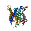
Spodoptera frugiperda (ツマジロクサヨトウ)
参照: UniProt: P47863
#2: 化合物
ChemComp-PEE / 1,2-dioleoyl-sn-glycero-3-phosphoethanolamine / DOPE


分子量: 744.034 Da / 分子数: 5 / 由来タイプ: 合成 / : C41H78NO8P / コメント: DOPE, リン脂質*YM
#3: 水 ChemComp-HOH / water


分子量: 18.015 Da / 分子数: 14 / 由来タイプ: 天然 / : H2O

-
実験情報

-
実験

実験手法: 電子線結晶学 / 使用した結晶の数: 1
EM実験試料の集合状態: 2D ARRAY / 3次元再構成法: 電子線結晶学

-
試料調製

構成要素名称: aquaporin-4 S180D mutant / タイプ: COMPLEX
試料包埋: NO / シャドウイング: NO / 染色: NO / 凍結: YES
結晶化温度: 293 K / 手法: microdialysis / pH: 6
詳細: 10mM MES (pH 6.0), 75mM NaCl, 50mM MgCl2, 2mM DTT, 1% glycerol, MICRODIALYSIS, temperature 293K

-
データ収集

顕微鏡モデル: JEOL 3000SFF
電子銃電子線源: FIELD EMISSION GUN / 加速電圧: 300 kV / 照射モード: FLOOD BEAM
電子レンズモード: DIFFRACTION
撮影フィルム・検出器のモデル: GENERIC CCD
回折平均測定温度: 4.2 K
放射光源由来: ELECTRON MICROSCOPE / タイプ: OTHER / 波長: 2.8 Å
検出器タイプ: Tietz / 検出器: CCD / 日付: 2008年2月4日
放射プロトコル: SINGLE WAVELENGTH / 単色(M)・ラウエ(L): M / 散乱光タイプ: electron
放射波長波長: 2.8 Å / 相対比: 1
反射解像度: 2.8→16.89 Å / Num. all: 10057 / Num. obs: 8750 / % possible obs: 87 % / Observed criterion σ(F): 0 / Observed criterion σ(I): 0 / Rmerge(I) obs: 0.197 / Rsym value: 0.197
反射 シェル解像度: 2.8→2.87 Å / % possible all: 80.15

-
解析

ソフトウェア
名称バージョン分類
REFMAC5.2.0019精密化
MRCデータ収集
MRCデータ削減
MRCデータスケーリング
CNS位相決定
3次元再構成対称性のタイプ: 2D CRYSTAL
精密化構造決定の手法: 分子置換
開始モデル: 2D57
解像度: 2.8→10 Å / Cor.coef. Fo:Fc: 0.926 / Cor.coef. Fo:Fc free: 0.839 / SU B: 41.271 / SU ML: 0.355 / 交差検証法: THROUGHOUT / σ(F): 0 / σ(I): 0 / ESU R Free: 0.395 / 立体化学のターゲット値: MAXIMUM LIKELIHOOD / 詳細: HYDROGENS HAVE BEEN ADDED IN THE RIDING POSITIONS
Rfactor反射数%反射Selection details
Rfree0.28788 481 4.9 %RANDOM
Rwork0.23079 ---
all0.234 9879 --
obs0.234 8089 81.88 %-
溶媒の処理イオンプローブ半径: 0.8 Å / 減衰半径: 0.8 Å / VDWプローブ半径: 1.2 Å / 溶媒モデル: MASK
原子変位パラメータBiso mean: 52.335 Å2
Baniso -1Baniso -2Baniso -3
1--6.68 Å20 Å20 Å2
2---6.68 Å20 Å2
3---13.37 Å2
精密化ステップサイクル: LAST / 解像度: 2.8→10 Å
タンパク質核酸リガンド溶媒全体
原子数1642 0 211 14 1867
拘束条件
Refine-IDタイプDev idealDev ideal target
ELECTRON CRYSTALLOGRAPHYr_bond_refined_d0.0110.0221889
ELECTRON CRYSTALLOGRAPHYr_angle_refined_deg1.5242.0272522
ELECTRON CRYSTALLOGRAPHYr_dihedral_angle_1_deg6.2975221
ELECTRON CRYSTALLOGRAPHYr_dihedral_angle_2_deg31.61523.01953
ELECTRON CRYSTALLOGRAPHYr_dihedral_angle_3_deg21.86815244
ELECTRON CRYSTALLOGRAPHYr_dihedral_angle_4_deg18.107153
ELECTRON CRYSTALLOGRAPHYr_chiral_restr0.090.2279
ELECTRON CRYSTALLOGRAPHYr_gen_planes_refined0.0040.021282
ELECTRON CRYSTALLOGRAPHYr_nbd_refined0.2610.21154
ELECTRON CRYSTALLOGRAPHYr_nbtor_refined0.3210.21286
ELECTRON CRYSTALLOGRAPHYr_xyhbond_nbd_refined0.1790.272
ELECTRON CRYSTALLOGRAPHYr_symmetry_vdw_refined0.2510.2107
ELECTRON CRYSTALLOGRAPHYr_symmetry_hbond_refined0.1020.23
ELECTRON CRYSTALLOGRAPHYr_mcbond_it0.6531.51113
ELECTRON CRYSTALLOGRAPHYr_mcangle_it1.15421749
ELECTRON CRYSTALLOGRAPHYr_scbond_it0.9153869
ELECTRON CRYSTALLOGRAPHYr_scangle_it1.5324.5773
ELECTRON CRYSTALLOGRAPHYr_rigid_bond_restr0.73131982
ELECTRON CRYSTALLOGRAPHYr_sphericity_free4.147314
ELECTRON CRYSTALLOGRAPHYr_sphericity_bonded0.77731853
LS精密化 シェル解像度: 2.8→2.867 Å / Total num. of bins used: 20
Rfactor反射数%反射
Rfree0.326 31 -
Rwork0.337 518 -
obs--80.15 %

+
万見について

-
お知らせ

-
2022年2月9日: EMDBエントリの付随情報ファイルのフォーマットが新しくなりました

EMDBエントリの付随情報ファイルのフォーマットが新しくなりました

  • EMDBのヘッダファイルのバージョン3が、公式のフォーマットとなりました。
  • これまでは公式だったバージョン1.9は、アーカイブから削除されます。

関連情報:EMDBヘッダ

外部リンク:wwPDBはEMDBデータモデルのバージョン3へ移行します

-
2020年8月12日: 新型コロナ情報

新型コロナ情報

URL: https://pdbjlvh1.pdbj.org/emnavi/covid19.php

新ページ: EM Navigatorに新型コロナウイルスの特設ページを開設しました。

関連情報:Covid-19情報 / 2020年3月5日: 新型コロナウイルスの構造データ

+
2020年3月5日: 新型コロナウイルスの構造データ

新型コロナウイルスの構造データ

関連情報:万見生物種 / 2020年8月12日: 新型コロナ情報

外部リンク:COVID-19特集ページ - PDBj / 今月の分子2020年2月:コロナウイルスプロテーアーゼ

+
2019年1月31日: EMDBのIDの桁数の変更

EMDBのIDの桁数の変更

  • EMDBエントリに付与されているアクセスコード(EMDB-ID)は4桁の数字(例、EMD-1234)でしたが、間もなく枯渇します。これまでの4桁のID番号は4桁のまま変更されませんが、4桁の数字を使い切った後に発行されるIDは5桁以上の数字(例、EMD-12345)になります。5桁のIDは2019年の春頃から発行される見通しです。
  • EM Navigator/万見では、接頭語「EMD-」は省略されています。

関連情報:Q: 「EMD」とは何ですか? / 万見/EM NavigatorにおけるID/アクセスコードの表記

外部リンク:EMDB Accession Codes are Changing Soon! / PDBjへお問い合わせ

+
2017年7月12日: PDB大規模アップデート

PDB大規模アップデート

  • 新バージョンのPDBx/mmCIF辞書形式に基づくデータがリリースされました。
  • 今回の更新はバージョン番号が4から5になる大規模なもので、全エントリデータの書き換えが行われる「Remediation」というアップデートに該当します。
  • このバージョンアップで、電子顕微鏡の実験手法に関する多くの項目の書式が改定されました(例:em_softwareなど)。
  • EM NavigatorとYorodumiでも、この改定に基づいた表示内容になります。

外部リンク:wwPDB Remediation / OneDepデータ基準に準拠した、より強化された内容のモデル構造ファイルが、PDBアーカイブで公開されました。

-
万見 (Yorodumi)

幾万の構造データを、幾万の視点から

  • 万見(Yorodumi)は、EMDB/PDB/SASBDBなどの構造データを閲覧するためのページです。
  • EM Navigatorの詳細ページの後継、Omokage検索のフロントエンドも兼ねています。

関連情報:EMDB / PDB / SASBDB / 3つのデータバンクの比較 / 万見検索 / 2016年8月31日: 新しいEM Navigatorと万見 / 万見文献 / Jmol/JSmol / 機能・相同性情報 / 新しいEM Navigatorと万見の変更点

他の情報も見る