 Movie
Movie Controller

[English] 日本語
 Yorodumi
- PDB-3iyz: Structure of Aquaporin-4 S180D mutant at 10.0 A resolution from e... -
+ Open data

- Basic information
| Entry | Database: PDB / ID: 3iyz | ||||||
|---|---|---|---|---|---|---|---|
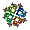
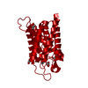
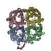
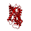
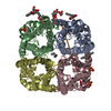
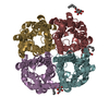
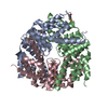
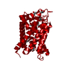
| Title | Structure of Aquaporin-4 S180D mutant at 10.0 A resolution from electron micrograph | ||||||
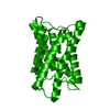
 Components Components | Aquaporin-4 | ||||||
 Keywords Keywords | TRANSPORT PROTEIN / WATER TRANSPORT / WATER CHANNEL / AQUAPORIN / TWO-DIMENSIONAL CRYSTAL / MEMBRANE PROTEIN / BACULOVIRUS EXPRESSION SYSTEM / GLYCOPROTEIN / MEMBRANE / PHOSPHOPROTEIN / TRANSMEMBRANE / TRANSPORT | ||||||
| Function / homology |  Function and homology information Function and homology informationPassive transport by Aquaporins / regulation of vascular endothelial growth factor production / cerebrospinal fluid secretion / cerebrospinal fluid circulation / astrocyte end-foot / intracellular water homeostasis / renal water absorption / negative regulation of cell adhesion molecule production / water channel activity / cell projection membrane ...Passive transport by Aquaporins / regulation of vascular endothelial growth factor production / cerebrospinal fluid secretion / cerebrospinal fluid circulation / astrocyte end-foot / intracellular water homeostasis / renal water absorption / negative regulation of cell adhesion molecule production / water channel activity / cell projection membrane / multicellular organismal-level water homeostasis / water transport / cellular response to interleukin-6 / Vasopressin regulates renal water homeostasis via Aquaporins / negative regulation of interleukin-1 beta production / negative regulation of interleukin-6 production / cellular response to interleukin-1 / T-tubule / establishment of localization in cell / basal plasma membrane / response to glucocorticoid / cellular response to estradiol stimulus / carbon dioxide transport / female pregnancy / cellular response to glucose stimulus / sensory perception of sound / cell-cell adhesion / sarcolemma / cellular response to type II interferon / cell-cell junction / protein homotetramerization / basolateral plasma membrane / response to ethanol / endosome membrane / external side of plasma membrane / protein-containing complex / extracellular region / identical protein binding / plasma membrane / cytoplasm Similarity search - Function | ||||||
| Biological species |  | ||||||
| Method | ELECTRON CRYSTALLOGRAPHY / electron crystallography /  MOLECULAR REPLACEMENT / cryo EM / Resolution: 10 Å MOLECULAR REPLACEMENT / cryo EM / Resolution: 10 Å | ||||||
 Authors Authors | Mitsuma, T. / Tani, K. / Hiroaki, Y. / Kamegawa, A. / Suzuki, H. / Hibino, H. / Kurachi, Y. / Fujiyoshi, Y. | ||||||
 Citation Citation | Journal: J Mol Biol / Year: 2010 Title: Influence of the cytoplasmic domains of aquaporin-4 on water conduction and array formation. Authors: Tadanori Mitsuma / Kazutoshi Tani / Yoko Hiroaki / Akiko Kamegawa / Hiroshi Suzuki / Hiroshi Hibino / Yoshihisa Kurachi / Yoshinori Fujiyoshi /  Abstract: Phosphorylation of Ser180 in cytoplasmic loop D has been shown to reduce the water permeability of aquaporin (AQP) 4, the predominant water channel in the brain. However, when the structure of the ...Phosphorylation of Ser180 in cytoplasmic loop D has been shown to reduce the water permeability of aquaporin (AQP) 4, the predominant water channel in the brain. However, when the structure of the S180D mutant (AQP4M23S180D), which was generated to mimic phosphorylated Ser180, was determined to 2.8 Å resolution using electron diffraction patterns, it showed no significant differences from the structure of the wild-type channel. High-resolution density maps usually do not resolve protein regions that are only partially ordered, but these can sometimes be seen in lower-resolution density maps calculated from electron micrographs. We therefore used images of two-dimensional crystals and determined the structure of AQP4M23S180D at 10 A resolution. The features of the 10-A density map are consistent with those of the previously determined atomic model; in particular, there were no indications of any obstruction near the cytoplasmic pore entrance. In addition, water conductance measurements, both in vitro and in vivo, show the same water permeability for wild-type and mutant AQP4M23, suggesting that the S180D mutation neither reduces water conduction through a conformational change nor reduces water conduction by interacting with a protein that would obstruct the cytoplasmic channel entrance. Finally, the 10-A map shows a cytoplasmic density in between four adjacent tetramers that most likely represents the association of four N termini. This finding supports the critical role of the N terminus of AQP4 in the stabilization of orthogonal arrays, as well as their interference through lipid modification of cysteine residues in the longer N-terminal isoform. | ||||||
| History |
|
- Structure visualization
| Movie |
Movie viewer |
|---|---|
| Structure viewer | Molecule: MolmilJmol/JSmol |
- Downloads & links
-Download
| PDBx/mmCIF format | 3iyz.cif.gz | 23.3 KB | Display | PDBx/mmCIF format |
|---|---|---|---|---|
| PDB format | pdb3iyz.ent.gz | 8.9 KB | Display | PDB format |
| PDBx/mmJSON format | 3iyz.json.gz | Tree view | PDBx/mmJSON format | |
| Others |  Other downloads Other downloads |
-Validation report
| Arichive directory | https://data.pdbj.org/pub/pdb/validation_reports/iy/3iyzftp://data.pdbj.org/pub/pdb/validation_reports/iy/3iyz | HTTPS FTP |
|---|
-Related structure data

-Links
 PDBj
PDBj


- Assembly
| Deposited unit | 
| ||||||||
|---|---|---|---|---|---|---|---|---|---|
| 1 | 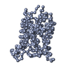
| ||||||||
| Unit cell |
|
-Components
| #1: Protein | Mass: 36595.137 Da / Num. of mol.: 1 / Fragment: UNP RESIDUES 23-323 / Mutation: YES Source method: isolated from a genetically manipulated source Source: (gene. exp.)   SPODOPTERA FRUGIPERDA (fall armyworm) / References: UniProt: P47863 SPODOPTERA FRUGIPERDA (fall armyworm) / References: UniProt: P47863 |
|---|
-Experimental details
-Experiment
| Experiment | Method: ELECTRON CRYSTALLOGRAPHY / Number of used crystals: 1 |
|---|---|
| EM experiment | Aggregation state: 2D ARRAY / 3D reconstruction method: electron crystallography |
- Sample preparation
| Component | Name: rat aquaporin-4 S180D mutant / Type: COMPLEX / Details: tetramer. The sample was embedded into lipid. |
|---|---|
| Molecular weight | Value: 0.032 MDa / Experimental value: NO |
| Buffer solution | Name: MES buffer / pH: 6 Details: 10mM MES, 75mM NaCl, 50mM MgCl2, 2mM DTT, 1% glycerol, 7% trehalose |
| Specimen | Embedding applied: NO / Shadowing applied: NO / Staining applied: NO / Vitrification applied: YES Details: 10mM MES, 75mM NaCl, 50mM MgCl2, 2mM DTT, 1% glycerol, 7% trehalose |
| Specimen support | Details: The molybdenum grids were covered with solid carbon |
| Vitrification | Instrument: REICHERT-JUNG PLUNGER / Cryogen name: NITROGEN / Chamber temperature: 277 K Method: The grid was blotted with filter paper and plunged into liquid nitrogen. |
| Crystal grow | pH: 6 / Details: pH 6.00 |
-Data collection
| Microscopy | Model: JEOL KYOTO-3000SFF |
|---|---|
| Electron gun | Electron source: FIELD EMISSION GUN / Accelerating voltage: 300 kV / Illumination mode: FLOOD BEAM |
| Electron lens | Mode: BRIGHT FIELD / Nominal magnification: 90000 X / Nominal defocus max: 3730 nm / Nominal defocus min: 430 nm / Cs: 1.6 mm Astigmatism: bjective lens astigmatism was corrected at 400,000 times magnification Camera length: 0 mm |
| Specimen holder | Specimen holder model: OTHER Specimen holder type: Top entry liquid helium cooled cryo specimen holder Temperature: 4.2 K / Tilt angle max: 60 ° / Tilt angle min: -60 ° |
| Image recording | Electron dose: 38 e/Å2 / Film or detector model: GENERIC CCD / Details: Tietz 4K CCD |
| Diffraction | Mean temperature: 4.2 K |
| Detector | Date: Feb 4, 2008 |
| Radiation | Protocol: SINGLE WAVELENGTH / Monochromatic (M) / Laue (L): M / Scattering type: electron |
| Radiation wavelength | Relative weight: 1 |
- Processing
| Software | Name: MRC / Classification: data scaling | ||||||||||||
|---|---|---|---|---|---|---|---|---|---|---|---|---|---|
| EM software |
| ||||||||||||
| CTF correction | Details: Each image | ||||||||||||
| 3D reconstruction | Method: 2D-crystals / Resolution: 10 Å / Resolution method: OTHER / Nominal pixel size: 1.67 Å / Magnification calibration: Each image / Details: MRC / Symmetry type: 2D CRYSTAL | ||||||||||||
| Atomic model building | Protocol: RIGID BODY FIT / Space: REAL / Target criteria: Cross-correlation coefficient / Details: REFINEMENT PROTOCOL--rigid body | ||||||||||||
| Atomic model building | PDB-ID: 2ZZ9 | ||||||||||||
| Refinement | Method to determine structure: MOLECULAR REPLACEMENT Starting model: PDB entry 2ZZ9 Highest resolution: 10 Å | ||||||||||||
| Refinement step | Cycle: LAST / Highest resolution: 10 Å
|
