 Movie
Movie Controller

[English] 日本語
 Yorodumi
- PDB-2f2b: Crystal structure of integral membrane protein Aquaporin AqpM at ... -
+ Open data

- Basic information
| Entry | Database: PDB / ID: 2f2b | ||||||
|---|---|---|---|---|---|---|---|
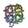
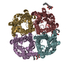
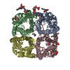
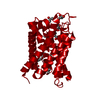
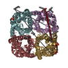
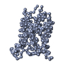
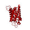
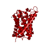
| Title | Crystal structure of integral membrane protein Aquaporin AqpM at 1.68A resolution | ||||||
 Components Components | Aquaporin aqpM | ||||||
 Keywords Keywords | MEMBRANE PROTEIN / protein / integral membrane protein / channel / Structural Genomics / PSI-2 / Protein Structure Initiative / Center for Structures of Membrane Proteins / CSMP | ||||||
| Function / homology |  Function and homology information Function and homology information | ||||||
| Biological species |   Methanothermobacter marburgensis str. Marburg (archaea) Methanothermobacter marburgensis str. Marburg (archaea) | ||||||
| Method |  X-RAY DIFFRACTION / SYNCHROTRON / MOLECULAR REPLACEMENT / Resolution: 1.68 Å X-RAY DIFFRACTION / SYNCHROTRON / MOLECULAR REPLACEMENT / Resolution: 1.68 Å | ||||||
 Authors Authors | Lee, J.K. / Kozono, D. / Remis, J. / Kitagawa, Y. / Agre, P. / Stroud, R.M. / Center for Structures of Membrane Proteins (CSMP) | ||||||
 Citation Citation | Journal: Proc.Natl.Acad.Sci.Usa / Year: 2005 Title: Structural basis for conductance by the archaeal aquaporin AqpM at 1.68 A. Authors: Lee, J.K. / Kozono, D. / Remis, J. / Kitagawa, Y. / Agre, P. / Stroud, R.M. | ||||||
| History |
|
- Structure visualization
| Structure viewer | Molecule: MolmilJmol/JSmol |
|---|
- Downloads & links
-Download
| PDBx/mmCIF format | 2f2b.cif.gz | 59.4 KB | Display | PDBx/mmCIF format |
|---|---|---|---|---|
| PDB format | pdb2f2b.ent.gz | 42.4 KB | Display | PDB format |
| PDBx/mmJSON format | 2f2b.json.gz | Tree view | PDBx/mmJSON format | |
| Others |  Other downloads Other downloads |
-Validation report
| Arichive directory | https://data.pdbj.org/pub/pdb/validation_reports/f2/2f2bftp://data.pdbj.org/pub/pdb/validation_reports/f2/2f2b | HTTPS FTP |
|---|
-Related structure data
| Related structure data |  2evuSC S: Starting model for refinement C: citing same article ( |
|---|---|
| Similar structure data |
-Links
 PDBj
PDBj
- Assembly
| Deposited unit | 
| ||||||||
|---|---|---|---|---|---|---|---|---|---|
| 1 | 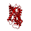
| ||||||||
| Unit cell |
| ||||||||
| Details | The biological assembly is a tetramer generated from the monomer in the asymmetric unit |
-Components
| #1: Protein | Mass: 25356.643 Da / Num. of mol.: 1 Source method: isolated from a genetically manipulated source Source: (gene. exp.) Methanothermobacter marburgensis str. Marburg (archaea)Species: Methanothermobacter marburgensis / Strain: Marburg / DSM 2133 / Gene: aqpM / Plasmid: pTRCa / Production host:  |
|---|---|
| #2: Chemical | ChemComp-GOL /   Mass: 92.094 Da / Num. of mol.: 1 / Source method: obtained synthetically / Formula: C3H8O3 Mass: 92.094 Da / Num. of mol.: 1 / Source method: obtained synthetically / Formula: C3H8O3 |
| #3: Water | ChemComp-HOH /  Mass: 18.015 Da / Num. of mol.: 129 / Source method: isolated from a natural source / Formula: H2O Mass: 18.015 Da / Num. of mol.: 129 / Source method: isolated from a natural source / Formula: H2O |
-Experimental details
-Experiment
| Experiment | Method: X-RAY DIFFRACTION / Number of used crystals: 1 |
|---|
- Sample preparation
| Crystal | Density Matthews: 3.09 Å3/Da / Density % sol: 60.15 % |
|---|---|
| Crystal grow | Temperature: 295 K / Method: vapor diffusion, hanging drop / pH: 8.5 Details: PEG4000 23%, 200 mM MgCl2, 10% glycerol, TRIS pH 8.5, VAPOR DIFFUSION, HANGING DROP, temperature 295K |
-Data collection
| Diffraction source | Source: SYNCHROTRON / Site: ALS  / Beamline: 8.3.1 / Wavelength: 1.1 Å / Beamline: 8.3.1 / Wavelength: 1.1 Å |
|---|---|
| Detector | Type: ADSC QUANTUM 4 / Detector: CCD / Date: Oct 15, 2005 / Details: KOHZU: Double Crystal Si(111) |
| Radiation | Monochromator: KOHZU: Double Crystal Si(111) / Protocol: SINGLE WAVELENGTH / Monochromatic (M) / Laue (L): M / Scattering type: x-ray |
| Radiation wavelength | Wavelength: 1.1 Å / Relative weight: 1 |
| Reflection | Resolution: 1.68→40 Å / Num. all: 35162 / Num. obs: 34031 / % possible obs: 96.8 % / Observed criterion σ(F): 2 / Observed criterion σ(I): 1 / Redundancy: 9.1 % / Biso Wilson estimate: 19.3 Å2 / Rmerge(I) obs: 0.078 / Net I/σ(I): 18.8 |
| Reflection shell | Resolution: 1.68→1.79 Å / Redundancy: 7.9 % / Rmerge(I) obs: 0.59 / Mean I/σ(I) obs: 3.3 / Num. unique all: 4576 / % possible all: 83.7 |
- Processing
| Software |
| |||||||||||||||||||||||||
|---|---|---|---|---|---|---|---|---|---|---|---|---|---|---|---|---|---|---|---|---|---|---|---|---|---|---|
| Refinement | Method to determine structure: MOLECULAR REPLACEMENT Starting model: pdb entry 2evu Resolution: 1.68→40 Å / Cross valid method: THROUGHOUT / σ(F): 0 / Stereochemistry target values: Engh & Huber
| |||||||||||||||||||||||||
| Refine analyze |
| |||||||||||||||||||||||||
| Refinement step | Cycle: LAST / Resolution: 1.68→40 Å
| |||||||||||||||||||||||||
| Refine LS restraints |
| |||||||||||||||||||||||||
| LS refinement shell | Resolution: 1.68→1.79 Å / Rfactor Rfree error: 0.018
|
