 Movie
Movie Controller

[English] 日本語
 Yorodumi
- PDB-2zi8: Crystal structure of the HsaC extradiol dioxygenase from M. tuber... -
+ Open data

- Basic information
| Entry | Database: PDB / ID: 2zi8 | ||||||
|---|---|---|---|---|---|---|---|
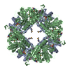
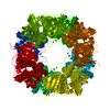
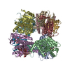
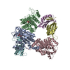
| Title | Crystal structure of the HsaC extradiol dioxygenase from M. tuberculosis in complex with 3,4-dihydroxy-9,10-seconandrost-1,3,5(10)-triene-9,17-dione (DHSA) | ||||||
 Components Components | PROBABLE BIPHENYL-2,3-DIOL 1,2-DIOXYGENASE BPHC | ||||||
 Keywords Keywords | OXIDOREDUCTASE / DHSA / HsaC / extradiol dioxygenase / Mycobacterium tuberculosis / Aromatic hydrocarbons catabolism / Iron | ||||||
| Function / homology |  Function and homology information Function and homology information3,4-dihydroxy-9,10-secoandrosta-1,3,5(10)-triene-9,17-dione 4,5-dioxygenase / 3,4-dihydroxy-9,10-secoandrosta-1,3,5(10)-triene-9,17-dione 4,5-dioxygenase activity / : / cholesterol catabolic process / response to cholesterol / cholesterol metabolic process / lipid catabolic process / ferrous iron binding / iron ion binding Similarity search - Function | ||||||
| Biological species |   Mycobacterium tuberculosis (bacteria) Mycobacterium tuberculosis (bacteria) | ||||||
| Method |  X-RAY DIFFRACTION / SYNCHROTRON / MOLECULAR REPLACEMENT / Resolution: 2.2 Å X-RAY DIFFRACTION / SYNCHROTRON / MOLECULAR REPLACEMENT / Resolution: 2.2 Å | ||||||
 Authors Authors | D'Angelo, I. / Yam, K.C. / Eltis, L.D. / Strynadka, N. | ||||||
 Citation Citation | Journal: Plos Pathog. / Year: 2009 Title: Studies of a ring-cleaving dioxygenase illuminate the role of cholesterol metabolism in the pathogenesis of Mycobacterium tuberculosis. Authors: Yam, K.C. / D'Angelo, I. / Kalscheuer, R. / Zhu, H. / Wang, J.X. / Snieckus, V. / Ly, L.H. / Converse, P.J. / Jacobs, W.R. / Strynadka, N. / Eltis, L.D. | ||||||
| History |
|
- Structure visualization
| Structure viewer | Molecule: MolmilJmol/JSmol |
|---|
- Downloads & links
-Download
| PDBx/mmCIF format | 2zi8.cif.gz | 142.8 KB | Display | PDBx/mmCIF format |
|---|---|---|---|---|
| PDB format | pdb2zi8.ent.gz | 111.3 KB | Display | PDB format |
| PDBx/mmJSON format | 2zi8.json.gz | Tree view | PDBx/mmJSON format | |
| Others |  Other downloads Other downloads |
-Validation report
| Arichive directory | https://data.pdbj.org/pub/pdb/validation_reports/zi/2zi8ftp://data.pdbj.org/pub/pdb/validation_reports/zi/2zi8 | HTTPS FTP |
|---|
-Related structure data
| Related structure data |  2zyqC 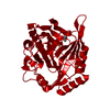 1hanS  2zf1 S: Starting model for refinement C: citing same article ( |
|---|---|
| Similar structure data |

-Links
 PDBj
PDBj


- Assembly
| Deposited unit | 
| ||||||||
|---|---|---|---|---|---|---|---|---|---|
| 1 | 
| ||||||||
| Unit cell |
|
-Components
| #1: Protein | Mass: 33628.312 Da / Num. of mol.: 2 Source method: isolated from a genetically manipulated source Source: (gene. exp.) Mycobacterium tuberculosis (bacteria) / Strain: H37Rv / Plasmid: pT7HC1 / Production host: References: UniProt: P96850, UniProt: P9WNW7*PLUS, biphenyl-2,3-diol 1,2-dioxygenase #2: Chemical |   Mass: 55.845 Da / Num. of mol.: 2 / Source method: obtained synthetically / Formula: Fe Mass: 55.845 Da / Num. of mol.: 2 / Source method: obtained synthetically / Formula: Fe#3: Chemical |   Mass: 316.391 Da / Num. of mol.: 2 / Source method: obtained synthetically / Formula: C19H24O4 Mass: 316.391 Da / Num. of mol.: 2 / Source method: obtained synthetically / Formula: C19H24O4#4: Water | ChemComp-HOH / |  Mass: 18.015 Da / Num. of mol.: 484 / Source method: isolated from a natural source / Formula: H2O Mass: 18.015 Da / Num. of mol.: 484 / Source method: isolated from a natural source / Formula: H2O |
|---|
-Experimental details
-Experiment
| Experiment | Method: X-RAY DIFFRACTION / Number of used crystals: 1 |
|---|
- Sample preparation
| Crystal | Density Matthews: 3.06 Å3/Da / Density % sol: 59.75 % |
|---|---|
| Crystal grow | Temperature: 291 K / Method: vapor diffusion, hanging drop / pH: 7 Details: 12-15% PEG 3350, 0.2M ammonium tartrate, 25% ethylene glycol, pH 7, VAPOR DIFFUSION, HANGING DROP, temperature 291K |
-Data collection
| Diffraction | Mean temperature: 100 K |
|---|---|
| Diffraction source | Source: SYNCHROTRON / Site: ALS  / Beamline: 8.2.2 / Wavelength: 1 Å / Beamline: 8.2.2 / Wavelength: 1 Å |
| Detector | Type: ADSC QUANTUM 315 / Detector: CCD / Date: Feb 20, 2007 / Details: MC |
| Radiation | Monochromator: Double crystal, Si(111) / Protocol: SINGLE WAVELENGTH / Monochromatic (M) / Laue (L): M / Scattering type: x-ray |
| Radiation wavelength | Wavelength: 1 Å / Relative weight: 1 |
| Reflection | Resolution: 2.2→30 Å / Num. all: 56012 / Num. obs: 55996 / % possible obs: 100 % / Observed criterion σ(F): 1 / Observed criterion σ(I): 1 / Rmerge(I) obs: 0.081 / Rsym value: 0.081 / Net I/σ(I): 26 |
| Reflection shell | Resolution: 2.2→2.32 Å / Rmerge(I) obs: 0.389 / Mean I/σ(I) obs: 6.1 / Num. unique all: 6156 / Rsym value: 0.389 / % possible all: 100 |
- Processing
| Software |
| |||||||||||||||||||||||||||||||||||||||||||||||||||||||||||||||||||||||||||||||||||||||||||||||
|---|---|---|---|---|---|---|---|---|---|---|---|---|---|---|---|---|---|---|---|---|---|---|---|---|---|---|---|---|---|---|---|---|---|---|---|---|---|---|---|---|---|---|---|---|---|---|---|---|---|---|---|---|---|---|---|---|---|---|---|---|---|---|---|---|---|---|---|---|---|---|---|---|---|---|---|---|---|---|---|---|---|---|---|---|---|---|---|---|---|---|---|---|---|---|---|---|
| Refinement | Method to determine structure: MOLECULAR REPLACEMENT Starting model: 1HAN Resolution: 2.2→20 Å / Cor.coef. Fo:Fc: 0.943 / Cor.coef. Fo:Fc free: 0.884 / SU B: 6.703 / SU ML: 0.166 / Cross valid method: THROUGHOUT / σ(F): 0 / σ(I): 0 / ESU R: 0.224 / ESU R Free: 0.212 / Stereochemistry target values: MAXIMUM LIKELIHOOD / Details: HYDROGENS HAVE BEEN ADDED IN THE RIDING POSITIONS
| |||||||||||||||||||||||||||||||||||||||||||||||||||||||||||||||||||||||||||||||||||||||||||||||
| Solvent computation | Ion probe radii: 0.8 Å / Shrinkage radii: 0.8 Å / VDW probe radii: 1.4 Å / Solvent model: MASK | |||||||||||||||||||||||||||||||||||||||||||||||||||||||||||||||||||||||||||||||||||||||||||||||
| Displacement parameters | Biso mean: 25.477 Å2
| |||||||||||||||||||||||||||||||||||||||||||||||||||||||||||||||||||||||||||||||||||||||||||||||
| Refinement step | Cycle: LAST / Resolution: 2.2→20 Å
| |||||||||||||||||||||||||||||||||||||||||||||||||||||||||||||||||||||||||||||||||||||||||||||||
| Refine LS restraints |
| |||||||||||||||||||||||||||||||||||||||||||||||||||||||||||||||||||||||||||||||||||||||||||||||
| LS refinement shell | Resolution: 2.2→2.257 Å / Total num. of bins used: 20
|
