[English] 日本語
Yorodumi
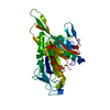
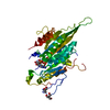
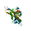
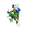
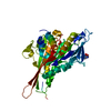
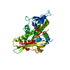
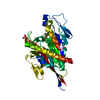
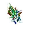
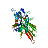
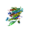
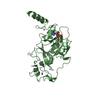
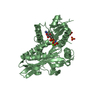
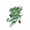
- PDB-2zfm: Crystal Structure of the Kif1A Motor Domain After Mg Release -

+
Open data


ID or keywords:

Loading...

-
Basic information

Entry
Database: PDB / ID: 2zfm
TitleCrystal Structure of the Kif1A Motor Domain After Mg Release
ComponentsKinesin-like protein KIF1A, Kinesin heavy chain isoform 5C
KeywordsTRANSPORT PROTEIN / kinesin / Alpha and Beta Protein / Enzyme / ATPase / P-loop / Motor Protein / ATP-binding / Coiled coil / Microtubule / Nucleotide-binding
Function / homology
Function and homology information


distal axon / anterograde dendritic transport of messenger ribonucleoprotein complex / protein transport along microtubule / neuronal dense core vesicle membrane / interkinetic nuclear migration / dense core granule cytoskeletal transport / anterograde neuronal dense core vesicle transport / anterograde dendritic transport of neurotransmitter receptor complex / retrograde neuronal dense core vesicle transport / Kinesins ...distal axon / anterograde dendritic transport of messenger ribonucleoprotein complex / protein transport along microtubule / neuronal dense core vesicle membrane / interkinetic nuclear migration / dense core granule cytoskeletal transport / anterograde neuronal dense core vesicle transport / anterograde dendritic transport of neurotransmitter receptor complex / retrograde neuronal dense core vesicle transport / Kinesins / regulation of dendritic spine development / anterograde axonal protein transport / intracellular mRNA localization / apolipoprotein receptor binding / COPI-dependent Golgi-to-ER retrograde traffic / cytoskeleton-dependent intracellular transport / regulation of dendritic spine morphogenesis / anterograde axonal transport / plus-end-directed microtubule motor activity / motor neuron axon guidance / ciliary rootlet / postsynaptic cytosol / synaptic vesicle transport / kinesin complex / microtubule motor activity / microtubule-based movement / neuronal dense core vesicle / mRNA transport / axonal growth cone / axon cytoplasm / vesicle-mediated transport / dendrite cytoplasm / axon guidance / Hydrolases; Acting on acid anhydrides; Acting on acid anhydrides to facilitate cellular and subcellular movement / synaptic vesicle / presynapse / postsynapse / microtubule binding / microtubule / neuron projection / axon / neuronal cell body / dendrite / perinuclear region of cytoplasm / ATP hydrolysis activity / protein-containing complex / ATP binding / identical protein binding / cytoplasm
Similarity search - Function
Kinesin-like KIF1-type / Kinesin protein 1B / Kinesin-like / Kinesin protein / Kinesin-associated / Kinesin-associated / Kinesin motor domain / Kinesin / Kinesin-like protein / Forkhead associated domain ...Kinesin-like KIF1-type / Kinesin protein 1B / Kinesin-like / Kinesin protein / Kinesin-associated / Kinesin-associated / Kinesin motor domain / Kinesin / Kinesin-like protein / Forkhead associated domain / Forkhead-associated (FHA) domain profile. / FHA domain / Forkhead-associated (FHA) domain / Kinesin motor domain signature. / Kinesin motor domain, conserved site / Kinesin motor domain / Kinesin motor domain profile. / Kinesin motor, catalytic domain. ATPase. / Kinesin motor domain / SMAD/FHA domain superfamily / Kinesin motor domain superfamily / PH domain / PH domain profile. / Pleckstrin homology domain. / Pleckstrin homology domain / PH-like domain superfamily / P-loop containing nucleoside triphosphate hydrolase / 3-Layer(aba) Sandwich / Alpha Beta
Similarity search - Domain/homology
ADENOSINE-5'-DIPHOSPHATE / Kinesin heavy chain isoform 5C / Kinesin-like protein KIF1A
Similarity search - Component
Biological speciesMus musculus (house mouse)
MethodX-RAY DIFFRACTION / SYNCHROTRON / MOLECULAR REPLACEMENT / Resolution: 2.31 Å
AuthorsNitta, R. / Okada, Y. / Hirokawa, N.
CitationJournal: Nat.Struct.Mol.Biol. / Year: 2008
Title: Structural model for strain-dependent microtubule activation of Mg-ADP release from kinesin.
Authors: Nitta, R. / Okada, Y. / Hirokawa, N.
History
DepositionJan 8, 2008Deposition site: PDBJ / Processing site: PDBJ
Revision 1.0Sep 23, 2008Provider: repository / Type: Initial release
Revision 1.1Jul 13, 2011Group: Version format compliance
Revision 1.2Aug 23, 2017Group: Source and taxonomy / Category: entity_src_gen
Revision 1.3Nov 1, 2023Group: Data collection / Database references / Refinement description
Category: chem_comp_atom / chem_comp_bond ...chem_comp_atom / chem_comp_bond / database_2 / pdbx_initial_refinement_model / struct_ref_seq_dif
Item: _database_2.pdbx_DOI / _database_2.pdbx_database_accession / _struct_ref_seq_dif.details

-
Structure visualization

Structure viewerMolecule:
MolmilJmol/JSmol

Downloads & links

-
Assembly

Deposited unit
A: Kinesin-like protein KIF1A, Kinesin heavy chain isoform 5C
hetero molecules


Theoretical massNumber of molelcules
Total (without water)41,5552
Polymers41,1281
Non-polymers4271
Water1,78399
1


  • Idetical with deposited unit
  • defined by author&software
TypeNameSymmetry operationNumber
identity operation1_555x,y,z1
MethodPISA
Unit cell
Length a, b, c (Å)41.767, 55.784, 155.956
Angle α, β, γ (deg.)90.00, 90.00, 90.00
Int Tables number19
Space group name H-MP212121

-
Components

#1: Protein Kinesin-like protein KIF1A, Kinesin heavy chain isoform 5C / Axonal transporter of synaptic vesi cles / Kinesin heavy chain neuron-specifiC 2


Mass: 41128.215 Da / Num. of mol.: 1 / Fragment: KIF1A (residues 1-355), KIF5C (residues 329-334)
Source method: isolated from a genetically manipulated source
Details: FUSION PROTEIN COMPRISES RESIDUES 1-355 OF Kinesin-like protein KIF1A, AND RESIDUES 329-334 OF Kinesin heavy chain isoform 5C, AND C-TERMINAL TAIL WITH SEQUENCE HHHHH
Source: (gene. exp.) Mus musculus (house mouse) / Plasmid: PET21B / Production host: Escherichia coli (E. coli) / References: UniProt: P33173, UniProt: P28738
#2: Chemical ChemComp-ADP / ADENOSINE-5'-DIPHOSPHATE / Adenosine diphosphate


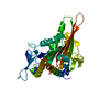
Mass: 427.201 Da / Num. of mol.: 1 / Source method: obtained synthetically / Formula: C10H15N5O10P2 / Comment: ADP, energy-carrying molecule*YM
#3: Water ChemComp-HOH / water / Water


Mass: 18.015 Da / Num. of mol.: 99 / Source method: isolated from a natural source / Formula: H2O

-
Experimental details

-
Experiment

ExperimentMethod: X-RAY DIFFRACTION

-
Sample preparation

CrystalDensity Matthews: 2.21 Å3/Da / Density % sol: 44.31 %
Crystal growTemperature: 293 K / Method: vapor diffusion, hanging drop / pH: 8.5
Details: pH 8.5, VAPOR DIFFUSION, HANGING DROP, temperature 293.0K

-
Data collection

DiffractionMean temperature: 93 K
Diffraction sourceSource: SYNCHROTRON / Site: Photon Factory / Beamline: BL-5A / Wavelength: 0.97 Å
DetectorType: ADSC QUANTUM 315 / Detector: CCD / Date: Feb 2, 2005
RadiationProtocol: SINGLE WAVELENGTH / Monochromatic (M) / Laue (L): M / Scattering type: x-ray
Radiation wavelengthWavelength: 0.97 Å / Relative weight: 1
ReflectionResolution: 2.3→50 Å / Num. obs: 16786 / Biso Wilson estimate: 33.1 Å2

-
Processing

Software
NameVersionClassification
CNS1refinement
ADSCQuantumdata collection
HKL-2000data reduction
HKL-2000data scaling
CNSphasing
RefinementMethod to determine structure: MOLECULAR REPLACEMENT
Starting model: 1I5S
Resolution: 2.31→33.44 Å / Rfactor Rfree error: 0.006 / Data cutoff high absF: 297463.31 / Data cutoff low absF: 0 / Isotropic thermal model: RESTRAINED / Cross valid method: THROUGHOUT / σ(F): 0
RfactorNum. reflection% reflectionSelection details
Rfree0.256 1558 10.1 %RANDOM
Rwork0.226 ---
obs0.226 15457 92 %-
Solvent computationSolvent model: FLAT MODEL / Bsol: 41.5287 Å2 / ksol: 0.334854 e/Å3
Displacement parametersBiso mean: 44.6 Å2
Baniso -1Baniso -2Baniso -3
1--6.38 Å20 Å20 Å2
2--22.18 Å20 Å2
3----15.8 Å2
Refine analyze
FreeObs
Luzzati coordinate error0.37 Å0.31 Å
Luzzati d res low-50 Å
Luzzati sigma a0.33 Å0.31 Å
Refinement stepCycle: LAST / Resolution: 2.31→33.44 Å
ProteinNucleic acidLigandSolventTotal
Num. atoms2579 0 27 99 2705
Refine LS restraints
Refine-IDTypeDev idealDev ideal target
X-RAY DIFFRACTIONc_bond_d0.01
X-RAY DIFFRACTIONc_angle_deg1.6
X-RAY DIFFRACTIONc_dihedral_angle_d25.9
X-RAY DIFFRACTIONc_improper_angle_d0.93
X-RAY DIFFRACTIONc_mcbond_it3.291.5
X-RAY DIFFRACTIONc_mcangle_it4.922
X-RAY DIFFRACTIONc_scbond_it5.232
X-RAY DIFFRACTIONc_scangle_it6.812.5
LS refinement shellResolution: 2.3→2.44 Å / Rfactor Rfree error: 0.023 / Total num. of bins used: 6
RfactorNum. reflection% reflection
Rfree0.333 206 9.2 %
Rwork0.306 2022 -
obs--80.7 %
Xplor file
Refine-IDSerial noParam fileTopol file
X-RAY DIFFRACTION1protein_rep.paramprotein.top
X-RAY DIFFRACTION2adp.paramadp.top
X-RAY DIFFRACTION3ion.paramion.top
X-RAY DIFFRACTION4water_rep.paramwater_rep.top

+
About Yorodumi

-
News

-
Feb 9, 2022. New format data for meta-information of EMDB entries

New format data for meta-information of EMDB entries

  • Version 3 of the EMDB header file is now the official format.
  • The previous official version 1.9 will be removed from the archive.

Related info.:EMDB header

External links:wwPDB to switch to version 3 of the EMDB data model

-
Aug 12, 2020. Covid-19 info

Covid-19 info

URL: https://pdbj.org/emnavi/covid19.php

New page: Covid-19 featured information page in EM Navigator.

Related info.:Covid-19 info / Mar 5, 2020. Novel coronavirus structure data

+
Mar 5, 2020. Novel coronavirus structure data

Novel coronavirus structure data

Related info.:Yorodumi Speices / Aug 12, 2020. Covid-19 info

External links:COVID-19 featured content - PDBj / Molecule of the Month (242):Coronavirus Proteases

+
Jan 31, 2019. EMDB accession codes are about to change! (news from PDBe EMDB page)

EMDB accession codes are about to change! (news from PDBe EMDB page)

  • The allocation of 4 digits for EMDB accession codes will soon come to an end. Whilst these codes will remain in use, new EMDB accession codes will include an additional digit and will expand incrementally as the available range of codes is exhausted. The current 4-digit format prefixed with “EMD-” (i.e. EMD-XXXX) will advance to a 5-digit format (i.e. EMD-XXXXX), and so on. It is currently estimated that the 4-digit codes will be depleted around Spring 2019, at which point the 5-digit format will come into force.
  • The EM Navigator/Yorodumi systems omit the EMD- prefix.

Related info.:Q: What is EMD? / ID/Accession-code notation in Yorodumi/EM Navigator

External links:EMDB Accession Codes are Changing Soon! / Contact to PDBj

+
Jul 12, 2017. Major update of PDB

Major update of PDB

  • wwPDB released updated PDB data conforming to the new PDBx/mmCIF dictionary.
  • This is a major update changing the version number from 4 to 5, and with Remediation, in which all the entries are updated.
  • In this update, many items about electron microscopy experimental information are reorganized (e.g. em_software).
  • Now, EM Navigator and Yorodumi are based on the updated data.

External links:wwPDB Remediation / Enriched Model Files Conforming to OneDep Data Standards Now Available in the PDB FTP Archive

-
Yorodumi

Thousand views of thousand structures

  • Yorodumi is a browser for structure data from EMDB, PDB, SASBDB, etc.
  • This page is also the successor to EM Navigator detail page, and also detail information page/front-end page for Omokage search.
  • The word "yorodu" (or yorozu) is an old Japanese word meaning "ten thousand". "mi" (miru) is to see.

Related info.:EMDB / PDB / SASBDB / Comparison of 3 databanks / Yorodumi Search / Aug 31, 2016. New EM Navigator & Yorodumi / Yorodumi Papers / Jmol/JSmol / Function and homology information / Changes in new EM Navigator and Yorodumi

Read more