 Movie
Movie Controller

[English] 日本語
 Yorodumi
- PDB-2zch: Crystal structure of human prostate specific antigen complexed wi... -
+ Open data

- Basic information
| Entry | Database: PDB / ID: 2zch | |||||||||
|---|---|---|---|---|---|---|---|---|---|---|
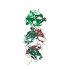
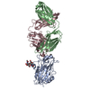
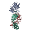
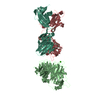
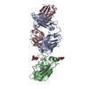
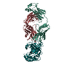
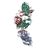
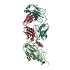
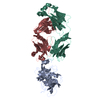
| Title | Crystal structure of human prostate specific antigen complexed with an activating antibody | |||||||||
 Components Components |
| |||||||||
 Keywords Keywords | IMMUNE SYSTEM / human PSA / kallikrein related peptidases / antibodies / prostate cancer / Glycoprotein / Hydrolase / Polymorphism / Protease / Secreted / Serine protease / Zymogen | |||||||||
| Function / homology |  Function and homology information Function and homology informationsemenogelase / positive regulation of antibacterial peptide production / hydrolase activity, acting on carbon-nitrogen (but not peptide) bonds, in linear amides / regulation of systemic arterial blood pressure / zymogen activation / negative regulation of angiogenesis / secretory granule / serine-type peptidase activity / Activated PKN1 stimulates transcription of AR (androgen receptor) regulated genes KLK2 and KLK3 / Regulation of Insulin-like Growth Factor (IGF) transport and uptake by Insulin-like Growth Factor Binding Proteins (IGFBPs) ...semenogelase / positive regulation of antibacterial peptide production / hydrolase activity, acting on carbon-nitrogen (but not peptide) bonds, in linear amides / regulation of systemic arterial blood pressure / zymogen activation / negative regulation of angiogenesis / secretory granule / serine-type peptidase activity / Activated PKN1 stimulates transcription of AR (androgen receptor) regulated genes KLK2 and KLK3 / Regulation of Insulin-like Growth Factor (IGF) transport and uptake by Insulin-like Growth Factor Binding Proteins (IGFBPs) / endopeptidase activity / serine-type endopeptidase activity / protein-containing complex / proteolysis / : / extracellular exosome / extracellular region / nucleus Similarity search - Function | |||||||||
| Biological species |  Homo sapiens (human) Homo sapiens (human) | |||||||||
| Method |  X-RAY DIFFRACTION / SYNCHROTRON / MOLECULAR REPLACEMENT / Resolution: 2.83 Å X-RAY DIFFRACTION / SYNCHROTRON / MOLECULAR REPLACEMENT / Resolution: 2.83 Å | |||||||||
 Authors Authors | Menez, R. / Stura, E. / Jolivet-Reynaud, C. | |||||||||
 Citation Citation | Journal: J.Mol.Biol. / Year: 2008 Title: Crystal structure of a ternary complex between human prostate-specific antigen, its substrate acyl intermediate and an activating antibody Authors: Michel, S. / Muller, B.H. / Bossus, M. / Ducancel, F. / Jolivet-Reynaud, C. / Stura, E.A. | |||||||||
| History |
|
- Structure visualization
| Structure viewer | Molecule: MolmilJmol/JSmol |
|---|
- Downloads & links
-Download
| PDBx/mmCIF format | 2zch.cif.gz | 145.9 KB | Display | PDBx/mmCIF format |
|---|---|---|---|---|
| PDB format | pdb2zch.ent.gz | 111.6 KB | Display | PDB format |
| PDBx/mmJSON format | 2zch.json.gz | Tree view | PDBx/mmJSON format | |
| Others |  Other downloads Other downloads |
-Validation report
| Arichive directory | https://data.pdbj.org/pub/pdb/validation_reports/zc/2zchftp://data.pdbj.org/pub/pdb/validation_reports/zc/2zch | HTTPS FTP |
|---|
-Related structure data
| Related structure data |  2zckC 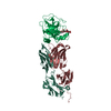 2zclC 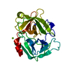 1l2eS 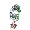 1rjlS C: citing same article ( S: Starting model for refinement |
|---|---|
| Similar structure data |


-Links
 PDBj
PDBj


- Assembly
| Deposited unit | 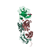
| ||||||||
|---|---|---|---|---|---|---|---|---|---|
| 1 |
| ||||||||
| Unit cell |
|
-Components
-Antibody , 2 types, 2 molecules LH
| #2: Antibody | Mass: 23444.730 Da / Num. of mol.: 1 / Source method: isolated from a natural source / Details: Ascitic fluids / Source: (natural) |
|---|---|
| #3: Antibody | Mass: 24366.205 Da / Num. of mol.: 1 / Source method: isolated from a natural source / Details: Ascitic fluids / Source: (natural) |
-Protein / Sugars , 2 types, 3 molecules P
| #1: Protein | Mass: 26122.025 Da / Num. of mol.: 1 / Fragment: UNP residues 25-261 / Source method: isolated from a natural source / Details: human seminal fluid / Source: (natural) Homo sapiens (human) / References: UniProt: P07288, semenogelase |
|---|---|
| #4: Sugar |  Type: D-saccharide, alpha linking / Mass: 221.208 Da / Num. of mol.: 2 Type: D-saccharide, alpha linking / Mass: 221.208 Da / Num. of mol.: 2Source method: isolated from a genetically manipulated source Formula: C8H15NO6 |
-Non-polymers , 3 types, 68 molecules 


| #5: Chemical | ChemComp-CL /  Mass: 35.453 Da / Num. of mol.: 1 / Source method: obtained synthetically / Formula: Cl Mass: 35.453 Da / Num. of mol.: 1 / Source method: obtained synthetically / Formula: Cl |
|---|---|
| #6: Chemical | ChemComp-PO4 /  Mass: 94.971 Da / Num. of mol.: 1 / Source method: obtained synthetically / Formula: PO4 Mass: 94.971 Da / Num. of mol.: 1 / Source method: obtained synthetically / Formula: PO4 |
| #7: Water | ChemComp-HOH / Mass: 18.015 Da / Num. of mol.: 66 / Source method: isolated from a natural source / Formula: H2O |
-Details
| Has protein modification | Y |
|---|---|
| Sequence details | A SEQUENCE DATABASE REFERENCE FOR CHAIN L AND H DOES NOT CURRENTLY EXIST. |
-Experimental details
-Experiment
| Experiment | Method: X-RAY DIFFRACTION / Number of used crystals: 1 |
|---|
- Sample preparation
| Crystal | Density Matthews: 3.05 Å3/Da / Density % sol: 59.66 % |
|---|---|
| Crystal grow | Temperature: 293 K / Method: vapor diffusion, sitting drop / pH: 7.2 Details: 14% MPEG 550, 100mM HEPES, pH7.2, VAPOR DIFFUSION, SITTING DROP, temperature 293K |
-Data collection
| Diffraction | Mean temperature: 200 K |
|---|---|
| Diffraction source | Source: SYNCHROTRON / Site: ESRF  / Beamline: ID23-2 / Wavelength: 0.873 Å / Beamline: ID23-2 / Wavelength: 0.873 Å |
| Detector | Type: MARMOSAIC 225 mm CCD / Detector: CCD / Date: Apr 21, 2006 |
| Radiation | Monochromator: horizontally side diffracting Silicon 111 crystal Protocol: SINGLE WAVELENGTH / Monochromatic (M) / Laue (L): M / Scattering type: x-ray |
| Radiation wavelength | Wavelength: 0.873 Å / Relative weight: 1 |
| Reflection | Resolution: 2.8→78.81 Å / Num. obs: 23506 / % possible obs: 100 % / Redundancy: 9.5 % / Rmerge(I) obs: 0.159 / Rsym value: 0.168 / Net I/σ(I): 14.5 |
| Reflection shell | Resolution: 2.8→2.95 Å / Redundancy: 9.7 % / Rmerge(I) obs: 0.498 / Mean I/σ(I) obs: 3.7 / Num. unique all: 3342 / Rsym value: 0.525 / % possible all: 100 |
- Processing
| Software |
| ||||||||||||||||||||||||||||||||||||||||||||||||||||||||||||||||||||||||||||||||||||||||||||||||||||||||||||||||||||||||||||||||||
|---|---|---|---|---|---|---|---|---|---|---|---|---|---|---|---|---|---|---|---|---|---|---|---|---|---|---|---|---|---|---|---|---|---|---|---|---|---|---|---|---|---|---|---|---|---|---|---|---|---|---|---|---|---|---|---|---|---|---|---|---|---|---|---|---|---|---|---|---|---|---|---|---|---|---|---|---|---|---|---|---|---|---|---|---|---|---|---|---|---|---|---|---|---|---|---|---|---|---|---|---|---|---|---|---|---|---|---|---|---|---|---|---|---|---|---|---|---|---|---|---|---|---|---|---|---|---|---|---|---|---|---|
| Refinement | Method to determine structure: MOLECULAR REPLACEMENT Starting model: PDB ENTRY 1L2E, 1RJL Resolution: 2.83→78.81 Å / Cor.coef. Fo:Fc: 0.924 / Cor.coef. Fo:Fc free: 0.856 / SU B: 13.127 / SU ML: 0.25 / Cross valid method: THROUGHOUT / ESU R: 1.996 / ESU R Free: 0.385 / Stereochemistry target values: MAXIMUM LIKELIHOOD / Details: HYDROGENS HAVE BEEN ADDED IN THE RIDING POSITIONS
| ||||||||||||||||||||||||||||||||||||||||||||||||||||||||||||||||||||||||||||||||||||||||||||||||||||||||||||||||||||||||||||||||||
| Solvent computation | Ion probe radii: 0.8 Å / Shrinkage radii: 0.8 Å / VDW probe radii: 1.2 Å / Solvent model: MASK | ||||||||||||||||||||||||||||||||||||||||||||||||||||||||||||||||||||||||||||||||||||||||||||||||||||||||||||||||||||||||||||||||||
| Displacement parameters | Biso mean: 38.776 Å2
| ||||||||||||||||||||||||||||||||||||||||||||||||||||||||||||||||||||||||||||||||||||||||||||||||||||||||||||||||||||||||||||||||||
| Refinement step | Cycle: LAST / Resolution: 2.83→78.81 Å
| ||||||||||||||||||||||||||||||||||||||||||||||||||||||||||||||||||||||||||||||||||||||||||||||||||||||||||||||||||||||||||||||||||
| Refine LS restraints |
| ||||||||||||||||||||||||||||||||||||||||||||||||||||||||||||||||||||||||||||||||||||||||||||||||||||||||||||||||||||||||||||||||||
| LS refinement shell | Resolution: 2.83→2.903 Å / Total num. of bins used: 20
|
