 Movie
Movie Controller


+ Open data

- Basic information
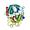
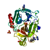
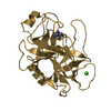
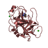
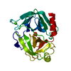
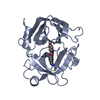
| Entry | Database: PDB / ID: 1l2e | ||||||
|---|---|---|---|---|---|---|---|
| Title | Human Kallikrein 6 (hK6) Active Form with benzamidine inhibitor | ||||||
 Components Components | Kallikrein 6 | ||||||
 Keywords Keywords | HYDROLASE / serine protease / kallikrein / human kallikrein 6 / benzamidine / protease / myelencephalon specific protease / zyme / protease M / neurosin | ||||||
| Function / homology |  Function and homology information Function and homology informationcornified envelope / tissue regeneration / positive regulation of G protein-coupled receptor signaling pathway / amyloid precursor protein metabolic process / hormone metabolic process / regulation of neuron projection development / protein autoprocessing / collagen catabolic process / regulation of cell differentiation / Hydrolases; Acting on peptide bonds (peptidases); Serine endopeptidases ...cornified envelope / tissue regeneration / positive regulation of G protein-coupled receptor signaling pathway / amyloid precursor protein metabolic process / hormone metabolic process / regulation of neuron projection development / protein autoprocessing / collagen catabolic process / regulation of cell differentiation / Hydrolases; Acting on peptide bonds (peptidases); Serine endopeptidases / intercellular bridge / myelination / secretory granule / central nervous system development / protein maturation / response to wounding / nuclear membrane / serine-type endopeptidase activity / nucleolus / mitochondrion / : / extracellular region / nucleoplasm / cytoplasm Similarity search - Function | ||||||
| Biological species |  Homo sapiens (human) Homo sapiens (human) | ||||||
| Method |  X-RAY DIFFRACTION / MOLECULAR REPLACEMENT / Resolution: 1.75 Å X-RAY DIFFRACTION / MOLECULAR REPLACEMENT / Resolution: 1.75 Å | ||||||
 Authors Authors | Bernett, M.J. / Blaber, S.I. / Scarisbrick, I.A. / Blaber, M. | ||||||
 Citation Citation | Journal: J.Biol.Chem. / Year: 2002 Title: Crystal structure and biochemical characterization of human kallikrein 6 reveals that a trypsin-like kallikrein is expressed in the central nervous system. Authors: Bernett, M.J. / Blaber, S.I. / Scarisbrick, I.A. / Dhanarajan, P. / Thompson, S.M. / Blaber, M. | ||||||
| History |
| ||||||
| Remark 999 | SEQUENCE The residue numbering is not sequential. |
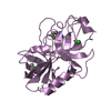
- Structure visualization
| Structure viewer | Molecule: MolmilJmol/JSmol |
|---|
- Downloads & links
-Download
| PDBx/mmCIF format | 1l2e.cif.gz | 59.6 KB | Display | PDBx/mmCIF format |
|---|---|---|---|---|
| PDB format | pdb1l2e.ent.gz | 42.5 KB | Display | PDB format |
| PDBx/mmJSON format | 1l2e.json.gz | Tree view | PDBx/mmJSON format | |
| Others |  Other downloads Other downloads |
-Validation report
| Arichive directory | https://data.pdbj.org/pub/pdb/validation_reports/l2/1l2eftp://data.pdbj.org/pub/pdb/validation_reports/l2/1l2e | HTTPS FTP |
|---|
-Related structure data
| Related structure data | 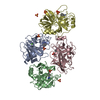 1a0jS S: Starting model for refinement |
|---|---|
| Similar structure data |


-Links
 PDBj
PDBj
- Assembly
| Deposited unit | 
| ||||||||
|---|---|---|---|---|---|---|---|---|---|
| 1 |
| ||||||||
| Unit cell |
|
-Components
| #1: Protein | Mass: 24532.877 Da / Num. of mol.: 1 Source method: isolated from a genetically manipulated source Source: (gene. exp.) Homo sapiens (human) / Cell line (production host): Sf9 / Production host:   Spodoptera frugiperda (fall armyworm) / References: UniProt: Q92876 Spodoptera frugiperda (fall armyworm) / References: UniProt: Q92876 |
|---|---|
| #2: Chemical | ChemComp-MG /   Mass: 24.305 Da / Num. of mol.: 1 / Source method: obtained synthetically / Formula: Mg Mass: 24.305 Da / Num. of mol.: 1 / Source method: obtained synthetically / Formula: Mg |
| #3: Chemical | ChemComp-BEN /   Mass: 120.152 Da / Num. of mol.: 1 / Source method: obtained synthetically / Formula: C7H8N2 Mass: 120.152 Da / Num. of mol.: 1 / Source method: obtained synthetically / Formula: C7H8N2 |
| #4: Water | ChemComp-HOH /  Mass: 18.015 Da / Num. of mol.: 139 / Source method: isolated from a natural source / Formula: H2O Mass: 18.015 Da / Num. of mol.: 139 / Source method: isolated from a natural source / Formula: H2O |
| Has protein modification | Y |
-Experimental details
-Experiment
| Experiment | Method: X-RAY DIFFRACTION / Number of used crystals: 1 |
|---|
- Sample preparation
| Crystal | Density Matthews: 2.12 Å3/Da / Density % sol: 42.01 % | ||||||||||||||||||||||||||||||||||||||||||||||||||||||||
|---|---|---|---|---|---|---|---|---|---|---|---|---|---|---|---|---|---|---|---|---|---|---|---|---|---|---|---|---|---|---|---|---|---|---|---|---|---|---|---|---|---|---|---|---|---|---|---|---|---|---|---|---|---|---|---|---|---|
| Crystal grow | Temperature: 277.15 K / Method: vapor diffusion, hanging drop / pH: 8.5 Details: 30% (w/v) PEG 4000, 0.2 M magnesium chloride hexahydrate, 0.1 M Tris hydrochloride, pH 8.5, VAPOR DIFFUSION, HANGING DROP, temperature 277.15K | ||||||||||||||||||||||||||||||||||||||||||||||||||||||||
| Crystal grow | *PLUS Temperature: 4 ℃ / pH: 4.5 | ||||||||||||||||||||||||||||||||||||||||||||||||||||||||
| Components of the solutions | *PLUS
|
-Data collection
| Diffraction | Mean temperature: 103 K |
|---|---|
| Diffraction source | Source: ROTATING ANODE / Type: RIGAKU / Wavelength: 1.5418 Å |
| Detector | Type: RIGAKU RAXIS IIC / Detector: IMAGE PLATE / Date: Oct 24, 2001 / Details: Osmic Blue Confocal Mirrors |
| Radiation | Monochromator: Osmic blue confocal mirrors / Protocol: SINGLE WAVELENGTH / Monochromatic (M) / Laue (L): M / Scattering type: x-ray |
| Radiation wavelength | Wavelength: 1.5418 Å / Relative weight: 1 |
| Reflection | Resolution: 1.75→43 Å / Num. all: 22918 / Num. obs: 21777 / % possible obs: 96 % / Observed criterion σ(F): 3 / Observed criterion σ(I): -3 / Redundancy: 21.6 % / Biso Wilson estimate: 26.6 Å2 / Rmerge(I) obs: 0.057 / Net I/σ(I): 43 |
| Reflection shell | Resolution: 1.75→1.79 Å / Rmerge(I) obs: 0.382 / Mean I/σ(I) obs: 4.9 / % possible all: 82.7 |
| Reflection | *PLUS Lowest resolution: 43 Å / Num. measured all: 495027 |
| Reflection shell | *PLUS % possible obs: 82.7 % |
- Processing
| Software |
| |||||||||||||||||||||||||
|---|---|---|---|---|---|---|---|---|---|---|---|---|---|---|---|---|---|---|---|---|---|---|---|---|---|---|
| Refinement | Method to determine structure: MOLECULAR REPLACEMENT Starting model: PDB ENTRY 1A0J Resolution: 1.75→43 Å / Cross valid method: THROUGHOUT / σ(F): 3
| |||||||||||||||||||||||||
| Displacement parameters | Biso mean: 26.6 Å2 | |||||||||||||||||||||||||
| Refinement step | Cycle: LAST / Resolution: 1.75→43 Å
| |||||||||||||||||||||||||
| Refine LS restraints |
| |||||||||||||||||||||||||
| LS refinement shell | Resolution: 1.75→1.86 Å
| |||||||||||||||||||||||||
| Refinement | *PLUS Lowest resolution: 43 Å | |||||||||||||||||||||||||
| Solvent computation | *PLUS | |||||||||||||||||||||||||
| Displacement parameters | *PLUS | |||||||||||||||||||||||||
| LS refinement shell | *PLUS Rfactor obs: 0.287 |
