 Movie
Movie Controller

[English] 日本語
 Yorodumi
- PDB-2za5: Crystal Structure of human tryptase with potent non-peptide inhibitor -
+ Open data

- Basic information
| Entry | Database: PDB / ID: 2za5 | ||||||
|---|---|---|---|---|---|---|---|
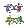
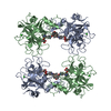
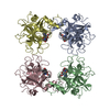
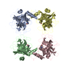
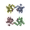
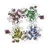
| Title | Crystal Structure of human tryptase with potent non-peptide inhibitor | ||||||
 Components Components | Tryptase beta 2 | ||||||
 Keywords Keywords | HYDROLASE / Tryptase / serine protease / tetramer | ||||||
| Function / homology |  Function and homology information Function and homology informationtryptase / Activation of Matrix Metalloproteinases / extracellular matrix disassembly / serine-type peptidase activity / defense response / extracellular matrix / serine-type endopeptidase activity / proteolysis / : / extracellular region / identical protein binding Similarity search - Function | ||||||
| Biological species |  Homo sapiens (human) Homo sapiens (human) | ||||||
| Method |  X-RAY DIFFRACTION / SYNCHROTRON / MOLECULAR REPLACEMENT / Resolution: 2.3 Å X-RAY DIFFRACTION / SYNCHROTRON / MOLECULAR REPLACEMENT / Resolution: 2.3 Å | ||||||
 Authors Authors | Spurlino, J.C. / Barnakov, S.A. / Lewandowski, F. / Milligan, C. | ||||||
 Citation Citation | Journal: Bioorg.Med.Chem.Lett. / Year: 2008 Title: Potent, nonpeptide inhibitors of human mast cell tryptase. Synthesis and biological evaluation of novel spirocyclic piperidine amide derivatives Authors: Costanzo, M.J. / Yabut, S.C. / Zhang, H.-C. / White, K.B. / de Garavilla, L. / Wang, Y. / Minor, L.K. / Tounge, B.A. / Barnakov, A.N. / Lewandowski, F. / Milligan, C. / Spurlino, J.C. / ...Authors: Costanzo, M.J. / Yabut, S.C. / Zhang, H.-C. / White, K.B. / de Garavilla, L. / Wang, Y. / Minor, L.K. / Tounge, B.A. / Barnakov, A.N. / Lewandowski, F. / Milligan, C. / Spurlino, J.C. / Abraham, W.M. / Boswell-Smith, V. / Page, C.P. / Maryanoff, B.E. #1: Journal: J.Med.Chem. / Year: 2003Title: Potent, small-molecule inhibitors of human mast cell tryptase. Antiasthmatic action of a dipeptide-based transition-state analogue containing a benzothiazole ketone Authors: Costanzo, M.J. / Yabut, S.C. / Almond, H.R. / Andrade-Gordon, P. / Corcoran, T.W. / de Garavilla, L. / Kauffman, J.A. / Abraham, W.M. / Recacha, R. / Chattopadhyay, D. / Maryanoff, B.E. | ||||||
| History |
|
- Structure visualization
| Structure viewer | Molecule: MolmilJmol/JSmol |
|---|
- Downloads & links
-Download
| PDBx/mmCIF format | 2za5.cif.gz | 209.4 KB | Display | PDBx/mmCIF format |
|---|---|---|---|---|
| PDB format | pdb2za5.ent.gz | 168.3 KB | Display | PDB format |
| PDBx/mmJSON format | 2za5.json.gz | Tree view | PDBx/mmJSON format | |
| Others |  Other downloads Other downloads |
-Validation report
| Arichive directory | https://data.pdbj.org/pub/pdb/validation_reports/za/2za5ftp://data.pdbj.org/pub/pdb/validation_reports/za/2za5 | HTTPS FTP |
|---|
-Related structure data
| Related structure data |  2zebC  2zecC  1a0lS S: Starting model for refinement C: citing same article ( |
|---|---|
| Similar structure data |

-Links
 PDBj
PDBj
- Assembly
| Deposited unit | 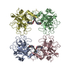
| ||||||||
|---|---|---|---|---|---|---|---|---|---|
| 1 |
| ||||||||
| Unit cell |
|
-Components
| #1: Protein | Mass: 27491.426 Da / Num. of mol.: 4 / Fragment: UNP residues 31-275 Source method: isolated from a genetically manipulated source Source: (gene. exp.) Homo sapiens (human) / Production host:  Pichia pastoris (fungus) / References: UniProt: Q6B052, UniProt: P20231*PLUS, tryptase Pichia pastoris (fungus) / References: UniProt: Q6B052, UniProt: P20231*PLUS, tryptase#2: Chemical | ChemComp-2FF / (   Mass: 412.480 Da / Num. of mol.: 4 / Source method: obtained synthetically / Formula: C26H24N2O3 Mass: 412.480 Da / Num. of mol.: 4 / Source method: obtained synthetically / Formula: C26H24N2O3#3: Water | ChemComp-HOH / |  Mass: 18.015 Da / Num. of mol.: 431 / Source method: isolated from a natural source / Formula: H2O Mass: 18.015 Da / Num. of mol.: 431 / Source method: isolated from a natural source / Formula: H2OHas protein modification | Y | |
|---|
-Experimental details
-Experiment
| Experiment | Method: X-RAY DIFFRACTION / Number of used crystals: 1 |
|---|
- Sample preparation
| Crystal | Density Matthews: 2.65 Å3/Da / Density % sol: 53.52 % |
|---|---|
| Crystal grow | Method: vapor diffusion, sitting drop / pH: 6.1 / Details: pH 6.1, VAPOR DIFFUSION, SITTING DROP |
-Data collection
| Diffraction | Mean temperature: 100 K |
|---|---|
| Diffraction source | Source: SYNCHROTRON / Site: APS  / Beamline: 17-ID / Wavelength: 1 Å / Beamline: 17-ID / Wavelength: 1 Å |
| Detector | Type: ADSC QUANTUM 210 / Detector: CCD |
| Radiation | Protocol: SINGLE WAVELENGTH / Monochromatic (M) / Laue (L): M / Scattering type: x-ray |
| Radiation wavelength | Wavelength: 1 Å / Relative weight: 1 |
| Reflection | Resolution: 2.3→20 Å / Num. obs: 51904 |
- Processing
| Software |
| ||||||||||||||||||||||||||||||||
|---|---|---|---|---|---|---|---|---|---|---|---|---|---|---|---|---|---|---|---|---|---|---|---|---|---|---|---|---|---|---|---|---|---|
| Refinement | Method to determine structure: MOLECULAR REPLACEMENT Starting model: PDB ENTRY 1A0L Resolution: 2.3→19.915 Å / FOM work R set: 0.839 / Cross valid method: THROUGHOUT
| ||||||||||||||||||||||||||||||||
| Solvent computation | Shrinkage radii: 0.9 Å / Solvent model: FLAT BULK SOLVENT MODEL / Bsol: 27.675 Å2 / ksol: 0.386 e/Å3 | ||||||||||||||||||||||||||||||||
| Displacement parameters | Biso max: 83.59 Å2 / Biso mean: 21.16 Å2 / Biso min: 5.49 Å2
| ||||||||||||||||||||||||||||||||
| Refinement step | Cycle: LAST / Resolution: 2.3→19.915 Å
| ||||||||||||||||||||||||||||||||
| Refine LS restraints |
|
