 Movie
Movie Controller

[English] 日本語
 Yorodumi
- PDB-2fxr: human beta tryptase II complexed with activated ketone inhibitor ... -
+ Open data

- Basic information
| Entry | Database: PDB / ID: 2fxr | ||||||
|---|---|---|---|---|---|---|---|
| Title | human beta tryptase II complexed with activated ketone inhibitor CRA-29382 | ||||||
 Components Components | Tryptase beta-2 | ||||||
 Keywords Keywords | HYDROLASE / serine protease / activated ketone inhibitor / pyrrolidine / CRA-29382 | ||||||
| Function / homology |  Function and homology information Function and homology informationtryptase / serine-type peptidase activity / extracellular matrix / serine-type endopeptidase activity / proteolysis / : Similarity search - Function | ||||||
| Biological species |  Homo sapiens (human) Homo sapiens (human) | ||||||
| Method |  X-RAY DIFFRACTION / SYNCHROTRON / MOLECULAR REPLACEMENT / Resolution: 2.5 Å X-RAY DIFFRACTION / SYNCHROTRON / MOLECULAR REPLACEMENT / Resolution: 2.5 Å | ||||||
 Authors Authors | Katz, B.A. | ||||||
 Citation Citation | Journal: Biochemistry / Year: 2006 Title: Structure-guided design of Peptide-based tryptase inhibitors. Authors: McGrath, M.E. / Sprengeler, P.A. / Hirschbein, B. / Somoza, J.R. / Lehoux, I. / Janc, J.W. / Gjerstad, E. / Graupe, M. / Estiarte, A. / Venkataramani, C. / Liu, Y. / Yee, R. / Ho, J.D. / ...Authors: McGrath, M.E. / Sprengeler, P.A. / Hirschbein, B. / Somoza, J.R. / Lehoux, I. / Janc, J.W. / Gjerstad, E. / Graupe, M. / Estiarte, A. / Venkataramani, C. / Liu, Y. / Yee, R. / Ho, J.D. / Green, M.J. / Lee, C.-S. / Liu, L. / Tai, V. / Spencer, J. / Sperandio, D. / Katz, B.A. | ||||||
| History |
|
- Structure visualization
| Structure viewer | Molecule: MolmilJmol/JSmol |
|---|
- Downloads & links
-Download
| PDBx/mmCIF format | 2fxr.cif.gz | 201.9 KB | Display | PDBx/mmCIF format |
|---|---|---|---|---|
| PDB format | pdb2fxr.ent.gz | 163.7 KB | Display | PDB format |
| PDBx/mmJSON format | 2fxr.json.gz | Tree view | PDBx/mmJSON format | |
| Others |  Other downloads Other downloads |
-Validation report
| Arichive directory | https://data.pdbj.org/pub/pdb/validation_reports/fx/2fxrftp://data.pdbj.org/pub/pdb/validation_reports/fx/2fxr | HTTPS FTP |
|---|
-Related structure data
| Related structure data | 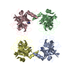 2fpzC  2fs8C  2fs9C  2fwwC 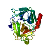 2fx4C 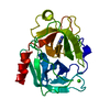 2fx6C  1a0lS C: citing same article ( S: Starting model for refinement |
|---|---|
| Similar structure data |


-Links
 PDBj
PDBj- Assembly
| Deposited unit | 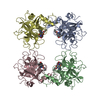
| ||||||||
|---|---|---|---|---|---|---|---|---|---|
| 1 |
| ||||||||
| Unit cell |
| ||||||||
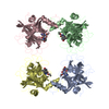
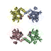
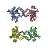
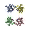
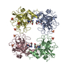
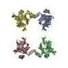
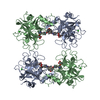
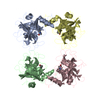
| Details | The biological unit is the tetramer. Each asymmetric unit contains one biologically-relevant tetramer. |
-Components
| #1: Protein | Mass: 27491.426 Da / Num. of mol.: 4 Source method: isolated from a genetically manipulated source Source: (gene. exp.) Homo sapiens (human) / Gene: TPSB2, TPS2 / Production host:  Pichia pastoris (fungus) / References: UniProt: P20231, tryptase Pichia pastoris (fungus) / References: UniProt: P20231, tryptase#2: Chemical | ChemComp-C3A / 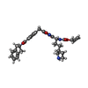  Mass: 557.640 Da / Num. of mol.: 4 / Source method: obtained synthetically / Formula: C31H35N5O5 Mass: 557.640 Da / Num. of mol.: 4 / Source method: obtained synthetically / Formula: C31H35N5O5#3: Water | ChemComp-HOH / |  Mass: 18.015 Da / Num. of mol.: 82 / Source method: isolated from a natural source / Formula: H2O Mass: 18.015 Da / Num. of mol.: 82 / Source method: isolated from a natural source / Formula: H2OHas protein modification | Y | |
|---|
-Experimental details
-Experiment
| Experiment | Method: X-RAY DIFFRACTION / Number of used crystals: 1 |
|---|
- Sample preparation
| Crystal | Density Matthews: 2.65 Å3/Da / Density % sol: 53.67 % |
|---|---|
| Crystal grow | Temperature: 293 K / Method: vapor diffusion, hanging drop Details: 2mg/mL protein, 10 mM MES, pH 6.1, 2M NaCl dissolved in 0.1 M NaOAc, pH 4.6, 0.2 M ammonium sulfate, 30% PEG 1500. Crystallization drops were set up using various ratios of protein solution ...Details: 2mg/mL protein, 10 mM MES, pH 6.1, 2M NaCl dissolved in 0.1 M NaOAc, pH 4.6, 0.2 M ammonium sulfate, 30% PEG 1500. Crystallization drops were set up using various ratios of protein solution to crystallization solution. Crystals appropriate for diffraction studies appeared in 2-5 days at room temperature, VAPOR DIFFUSION, HANGING DROP, temperature 293.0K |
-Data collection
| Diffraction | Mean temperature: 113 K |
|---|---|
| Diffraction source | Source: SYNCHROTRON / Site: ALS  / Beamline: 5.0.1 / Wavelength: 1 Å / Beamline: 5.0.1 / Wavelength: 1 Å |
| Detector | Type: ADSC QUANTUM 4 / Detector: CCD / Date: Oct 9, 2004 |
| Radiation | Protocol: SINGLE WAVELENGTH / Monochromatic (M) / Laue (L): M / Scattering type: x-ray |
| Radiation wavelength | Wavelength: 1 Å / Relative weight: 1 |
| Reflection | Resolution: 2.5→42.78 Å / Num. all: 39151 / Num. obs: 39151 / % possible obs: 99.9 % / Observed criterion σ(F): 0 / Observed criterion σ(I): 0 / Rmerge(I) obs: 0.101 |
| Reflection shell | Resolution: 2.5→2.54 Å / Rmerge(I) obs: 0.557 / % possible all: 98.5 |
- Processing
| Software |
| ||||||||||||||||||||
|---|---|---|---|---|---|---|---|---|---|---|---|---|---|---|---|---|---|---|---|---|---|
| Refinement | Method to determine structure: MOLECULAR REPLACEMENT Starting model: PDB ENTRY 1A0L Resolution: 2.5→42.78 Å / σ(F): 0 / Stereochemistry target values: Engh & Huber
| ||||||||||||||||||||
| Refinement step | Cycle: LAST / Resolution: 2.5→42.78 Å
| ||||||||||||||||||||
| Refine LS restraints |
| ||||||||||||||||||||
| Xplor file |
|
