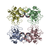
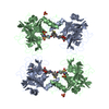
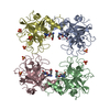
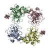
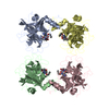
SHEET DETERMINATION METHOD: DSSP THE SHEETS PRESENTED AS "DB" IN EACH CHAIN ON SHEET RECORDS BELOW ... SHEET DETERMINATION METHOD: DSSP THE SHEETS PRESENTED AS "DB" IN EACH CHAIN ON SHEET RECORDS BELOW IS ACTUALLY AN 6-STRANDED BARREL THIS IS REPRESENTED BY A 7-STRANDED SHEET IN WHICH THE FIRST AND LAST STRANDS ARE IDENTICAL.
Protocol: SINGLE WAVELENGTH / Monochromatic (M) / Laue (L): M / Scattering type: x-ray
Radiation wavelength
Wavelength: 0.933 Å / Relative weight: 1
Reflection
Resolution: 2.04→58.72 Å / Num. obs: 62107 / % possible obs: 86.8 % / Observed criterion σ(I): 0 / Redundancy: 1.9 % / Biso Wilson estimate: 34.76 Å2 / Rmerge(I) obs: 0.07 / Net I/σ(I): 19.1
Reflection shell
Resolution: 2.04→2.15 Å / Redundancy: 2.1 % / Rmerge(I) obs: 0.29 / Mean I/σ(I) obs: 2.2 / % possible all: 81.8
-
Processing
Software
Name
Version
Classification
BUSTER
2.11.1
refinement
MOSFLM
datareduction
SCALA
datascaling
AMoRE
phasing
Refinement
Method to determine structure: MOLECULAR REPLACEMENT Starting model: IN-HOUSE STRUCTURE Resolution: 2.05→30.01 Å / Cor.coef. Fo:Fc: 0.9356 / Cor.coef. Fo:Fc free: 0.9224 / SU R Cruickshank DPI: 0.211 / Cross valid method: THROUGHOUT / σ(F): 0 / SU R Blow DPI: 0.206 / SU Rfree Blow DPI: 0.16 / SU Rfree Cruickshank DPI: 0.163 Details: THE P43 RESIDUES ARE THE INHIBITOR IN THE ACTIVE SITE.
Rfactor
Num. reflection
% reflection
Selection details
Rfree
0.2113
3140
5.07 %
RANDOM
Rwork
0.1884
-
-
-
obs
0.1896
61993
87.63 %
-
Displacement parameters
Biso mean: 32.72 Å2
Baniso -1
Baniso -2
Baniso -3
1-
3.3731 Å2
0 Å2
0 Å2
2-
-
3.3731 Å2
0 Å2
3-
-
-
-6.7463 Å2
Refine analyze
Luzzati coordinate error obs: 0.241 Å
Refinement step
Cycle: LAST / Resolution: 2.05→30.01 Å
Protein
Nucleic acid
Ligand
Solvent
Total
Num. atoms
7648
0
120
366
8134
Refine LS restraints
Refine-ID
Type
Dev ideal
Number
Restraint function
Weight
X-RAY DIFFRACTION
t_bond_d
0.01
8056
HARMONIC
2
X-RAY DIFFRACTION
t_angle_deg
1.09
11056
HARMONIC
2
X-RAY DIFFRACTION
t_dihedral_angle_d
2532
SINUSOIDAL
2
X-RAY DIFFRACTION
t_incorr_chiral_ct
X-RAY DIFFRACTION
t_pseud_angle
X-RAY DIFFRACTION
t_trig_c_planes
204
HARMONIC
2
X-RAY DIFFRACTION
t_gen_planes
1180
HARMONIC
5
X-RAY DIFFRACTION
t_it
8056
HARMONIC
20
X-RAY DIFFRACTION
t_nbd
X-RAY DIFFRACTION
t_omega_torsion
3.52
X-RAY DIFFRACTION
t_other_torsion
17.01
X-RAY DIFFRACTION
t_improper_torsion
X-RAY DIFFRACTION
t_chiral_improper_torsion
972
SEMIHARMONIC
5
X-RAY DIFFRACTION
t_sum_occupancies
X-RAY DIFFRACTION
t_utility_distance
X-RAY DIFFRACTION
t_utility_angle
X-RAY DIFFRACTION
t_utility_torsion
X-RAY DIFFRACTION
t_ideal_dist_contact
9157
SEMIHARMONIC
4
LS refinement shell
Resolution: 2.05→2.1 Å / Total num. of bins used: 20
Rfactor
Num. reflection
% reflection
Rfree
0.212
262
5.63 %
Rwork
0.1943
4391
-
all
0.1953
4653
-
obs
-
-
87.63 %
+
About Yorodumi
-
News
-
Feb 9, 2022. New format data for meta-information of EMDB entries
New format data for meta-information of EMDB entries
Version 3 of the EMDB header file is now the official format.
The previous official version 1.9 will be removed from the archive.
In the structure databanks used in Yorodumi, some data are registered as the other names, "COVID-19 virus" and "2019-nCoV". Here are the details of the virus and the list of structure data.
Jan 31, 2019. EMDB accession codes are about to change! (news from PDBe EMDB page)
EMDB accession codes are about to change! (news from PDBe EMDB page)
The allocation of 4 digits for EMDB accession codes will soon come to an end. Whilst these codes will remain in use, new EMDB accession codes will include an additional digit and will expand incrementally as the available range of codes is exhausted. The current 4-digit format prefixed with “EMD-” (i.e. EMD-XXXX) will advance to a 5-digit format (i.e. EMD-XXXXX), and so on. It is currently estimated that the 4-digit codes will be depleted around Spring 2019, at which point the 5-digit format will come into force.
The EM Navigator/Yorodumi systems omit the EMD- prefix.
Related info.:Q: What is EMD? / ID/Accession-code notation in Yorodumi/EM Navigator
Yorodumi is a browser for structure data from EMDB, PDB, SASBDB, etc.
This page is also the successor to EM Navigator detail page, and also detail information page/front-end page for Omokage search.
The word "yorodu" (or yorozu) is an old Japanese word meaning "ten thousand". "mi" (miru) is to see.
Related info.:EMDB / PDB / SASBDB / Comparison of 3 databanks / Yorodumi Search / Aug 31, 2016. New EM Navigator & Yorodumi / Yorodumi Papers / Jmol/JSmol / Function and homology information / Changes in new EM Navigator and Yorodumi
 Movie
Movie Controller


 Open data
 Basic information
 Components
 Keywords
 Function and homology information
 HOMO SAPIENS (human)
 X-RAY DIFFRACTION /
  Authors
 Citation
 Structure visualization
 Downloads & links
 Other downloads

 PDBj
PDBj
 Assembly
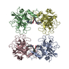
 PICHIA PASTORIS (fungus) / References: UniProt: Q15661, UniProt: P20231*PLUS, tryptase
 Mass: 402.461 Da / Num. of mol.: 4 / Source method: obtained synthetically / Formula: C25H23FN2O2
 Mass: 18.015 Da / Num. of mol.: 366 / Source method: isolated from a natural source / Formula: H2O
 Sample preparation
 / Beamline: ID14-2 / Wavelength: 0.933
  Processing