| Entry | Database: PDB / ID: 2z9i
|
|---|
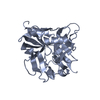
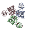
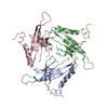
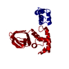
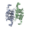
| Title | Crystal structure of RV0983 from Mycobacterium tuberculosis- Proteolytically active form |
|---|
 Components Components | - GATV
- PROBABLE SERINE PROTEASE PEPD
- SVEQV
|
|---|
 Keywords Keywords | HYDROLASE / SERINE PROTEASE / HtrA |
|---|
| Function / homology |  Function and homology information Function and homology information
peptidase Do / cellular response to antibiotic / serine-type peptidase activity / protein catabolic process / serine-type endopeptidase activity / proteolysis / extracellular region / plasma membraneSimilarity search - Function : / PDZ domain / Peptidase S1C / Trypsin-like peptidase domain / PDZ domain / Pdz3 Domain / PDZ domain profile. / Domain present in PSD-95, Dlg, and ZO-1/2. / PDZ domain / PDZ superfamily ...: / PDZ domain / Peptidase S1C / Trypsin-like peptidase domain / PDZ domain / Pdz3 Domain / PDZ domain profile. / Domain present in PSD-95, Dlg, and ZO-1/2. / PDZ domain / PDZ superfamily / Trypsin-like serine proteases / Thrombin, subunit H / Roll / Peptidase S1, PA clan, chymotrypsin-like fold / Peptidase S1, PA clan / Beta Barrel / Mainly BetaSimilarity search - Domain/homology |
|---|
| Biological species |   Mycobacterium tuberculosis (bacteria) Mycobacterium tuberculosis (bacteria) |
|---|
| Method |  X-RAY DIFFRACTION / SYNCHROTRON / MAD / Resolution: 2 Å X-RAY DIFFRACTION / SYNCHROTRON / MAD / Resolution: 2 Å |
|---|
 Authors Authors | Palaninathan, S.K. / Mohamedmohaideen, N.N. / Sacchettini, J.C. |
|---|
 Citation Citation | Journal: Biochemistry / Year: 2008
Title: Structure and function of the virulence-associated high-temperature requirement A of Mycobacterium tuberculosis
Authors: Mohamedmohaideen, N.N. / Palaninathan, S.K. / Morin, P.M. / Williams, B.J. / Braunstein, M. / Tichy, S.E. / Locker, J. / Russell, D.H. / Jacobs, W.R. / Sacchettini, J.C. |
|---|
| History | | Deposition | Sep 20, 2007 | Deposition site: PDBJ / Processing site: PDBJ |
|---|
| Revision 1.0 | Jun 10, 2008 | Provider: repository / Type: Initial release |
|---|
| Revision 1.1 | Jul 13, 2011 | Group: Version format compliance |
|---|
| Revision 1.2 | Oct 30, 2024 | Group: Data collection / Database references ...Data collection / Database references / Derived calculations / Refinement description / Structure summary
Category: chem_comp_atom / chem_comp_bond ...chem_comp_atom / chem_comp_bond / database_2 / pdbx_entry_details / pdbx_modification_feature / struct_conn / struct_ncs_dom_lim / struct_ref_seq_dif
Item: _database_2.pdbx_DOI / _database_2.pdbx_database_accession ..._database_2.pdbx_DOI / _database_2.pdbx_database_accession / _struct_conn.pdbx_dist_value / _struct_conn.pdbx_leaving_atom_flag / _struct_conn.ptnr1_auth_asym_id / _struct_conn.ptnr1_auth_comp_id / _struct_conn.ptnr1_auth_seq_id / _struct_conn.ptnr1_label_asym_id / _struct_conn.ptnr1_label_atom_id / _struct_conn.ptnr1_label_comp_id / _struct_conn.ptnr1_label_seq_id / _struct_conn.ptnr2_auth_asym_id / _struct_conn.ptnr2_auth_comp_id / _struct_conn.ptnr2_auth_seq_id / _struct_conn.ptnr2_label_asym_id / _struct_conn.ptnr2_label_atom_id / _struct_conn.ptnr2_label_comp_id / _struct_conn.ptnr2_label_seq_id / _struct_ncs_dom_lim.beg_auth_comp_id / _struct_ncs_dom_lim.end_auth_comp_id / _struct_ref_seq_dif.details |
|---|
|
|---|
 Movie
Movie Controller

 Yorodumi
 Open data
 Basic information
 Structure visualization
 Downloads & links
 Other downloads


 PDBj
PDBj
 Assembly