 Movie
Movie Controller


+ Open data

- Basic information
| Entry | Database: PDB / ID: 1ky9 | ||||||
|---|---|---|---|---|---|---|---|
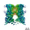
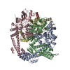
| Title | Crystal Structure of DegP (HtrA) | ||||||
 Components Components | PROTEASE DO | ||||||
 Keywords Keywords | HYDROLASE / protein quality control / serine protease / trypsin / chaperone / PDZ / ATP-independent / temperature-regulated / periplasm / cage-forming protein | ||||||
| Function / homology |  Function and homology information Function and homology informationpeptidase Do / response to temperature stimulus / Secretion of toxins / protein quality control for misfolded or incompletely synthesized proteins / serine-type peptidase activity / peptidase activity / outer membrane-bounded periplasmic space / response to heat / protein folding / response to oxidative stress ...peptidase Do / response to temperature stimulus / Secretion of toxins / protein quality control for misfolded or incompletely synthesized proteins / serine-type peptidase activity / peptidase activity / outer membrane-bounded periplasmic space / response to heat / protein folding / response to oxidative stress / periplasmic space / serine-type endopeptidase activity / proteolysis / identical protein binding / plasma membrane Similarity search - Function | ||||||
| Biological species |  | ||||||
| Method |  X-RAY DIFFRACTION / SYNCHROTRON / MAD / Resolution: 2.8 Å X-RAY DIFFRACTION / SYNCHROTRON / MAD / Resolution: 2.8 Å | ||||||
 Authors Authors | Krojer, T. / Garrido-Franco, M. / Huber, R. / Ehrmann, M. / Clausen, T. | ||||||
 Citation Citation | Journal: Nature / Year: 2002 Title: Crystal structure of DegP (HtrA) reveals a new protease-chaperone machine. Authors: Krojer, T. / Garrido-Franco, M. / Huber, R. / Ehrmann, M. / Clausen, T. | ||||||
| History |
|
- Structure visualization
| Structure viewer | Molecule: MolmilJmol/JSmol |
|---|
- Downloads & links
-Download
| PDBx/mmCIF format | 1ky9.cif.gz | 147.9 KB | Display | PDBx/mmCIF format |
|---|---|---|---|---|
| PDB format | pdb1ky9.ent.gz | 116.6 KB | Display | PDB format |
| PDBx/mmJSON format | 1ky9.json.gz | Tree view | PDBx/mmJSON format | |
| Others |  Other downloads Other downloads |
-Validation report
| Arichive directory | https://data.pdbj.org/pub/pdb/validation_reports/ky/1ky9ftp://data.pdbj.org/pub/pdb/validation_reports/ky/1ky9 | HTTPS FTP |
|---|
-Related structure data
| Similar structure data |
|---|
-Links
 PDBj
PDBj

- Assembly
| Deposited unit | 
| ||||||||
|---|---|---|---|---|---|---|---|---|---|
| 1 | x 6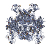
| ||||||||
| 2 | x 6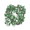
| ||||||||
| Unit cell |
| ||||||||
| Details | Hexamers of molecule A and B are entirely formed by crystal symmetry. |
-Components
| #1: Protein | Mass: 47509.449 Da / Num. of mol.: 2 / Mutation: S210A Source method: isolated from a genetically manipulated source Source: (gene. exp.) References: UniProt: P0C0V0, Hydrolases; Acting on peptide bonds (peptidases); Serine endopeptidases #2: Water | ChemComp-HOH / |  Mass: 18.015 Da / Num. of mol.: 166 / Source method: isolated from a natural source / Formula: H2O Mass: 18.015 Da / Num. of mol.: 166 / Source method: isolated from a natural source / Formula: H2OHas protein modification | Y | |
|---|
-Experimental details
-Experiment
| Experiment | Method: X-RAY DIFFRACTION / Number of used crystals: 1 |
|---|
- Sample preparation
| Crystal | Density Matthews: 2.61 Å3/Da / Density % sol: 52.94 % | ||||||||||||||||||||||||
|---|---|---|---|---|---|---|---|---|---|---|---|---|---|---|---|---|---|---|---|---|---|---|---|---|---|
| Crystal grow | Temperature: 291 K / Method: vapor diffusion, sitting drop / pH: 8.5 Details: Isopropanol, PEG 2000 MME, Tris, pH 8.5, VAPOR DIFFUSION, SITTING DROP, temperature 291K | ||||||||||||||||||||||||
| Crystal grow | *PLUS Temperature: 18 ℃ / Method: vapor diffusion | ||||||||||||||||||||||||
| Components of the solutions | *PLUS
|
-Data collection
| Diffraction | Mean temperature: 100 K | ||||||||||||
|---|---|---|---|---|---|---|---|---|---|---|---|---|---|
| Diffraction source | Source: SYNCHROTRON / Site: ESRF  / Beamline: ID14-4 / Wavelength: 0.9500, 0.9795, 0.9797 / Beamline: ID14-4 / Wavelength: 0.9500, 0.9795, 0.9797 | ||||||||||||
| Detector | Type: ADSC QUANTUM 4 / Detector: CCD / Date: Jun 22, 2001 | ||||||||||||
| Radiation | Monochromator: Si111 or Si311 crystals, LN2 cooled / Protocol: MAD / Monochromatic (M) / Laue (L): M / Scattering type: x-ray | ||||||||||||
| Radiation wavelength |
| ||||||||||||
| Reflection | Resolution: 2.8→20 Å / Num. all: 48422 / Num. obs: 48312 / % possible obs: 97 % / Observed criterion σ(F): -3 / Observed criterion σ(I): -3 / Redundancy: 4.4 % / Biso Wilson estimate: 82 Å2 / Rsym value: 0.083 | ||||||||||||
| Reflection shell | Resolution: 2.8→2.9 Å / Redundancy: 3.2 % / Mean I/σ(I) obs: 2.3 / Num. unique all: 4397 / Rsym value: 0.469 / % possible all: 92.2 | ||||||||||||
| Reflection | *PLUS Lowest resolution: 20 Å / Rmerge(I) obs: 0.083 | ||||||||||||
| Reflection shell | *PLUS % possible obs: 92.2 % / Rmerge(I) obs: 0.469 |
- Processing
| Software |
| ||||||||||||||||||||
|---|---|---|---|---|---|---|---|---|---|---|---|---|---|---|---|---|---|---|---|---|---|
| Refinement | Method to determine structure: MAD / Resolution: 2.8→20 Å / Cross valid method: THROUGHOUT / σ(F): 0 / Stereochemistry target values: Engh & Huber
| ||||||||||||||||||||
| Refinement step | Cycle: LAST / Resolution: 2.8→20 Å
| ||||||||||||||||||||
| Refine LS restraints |
| ||||||||||||||||||||
| Refinement | *PLUS Lowest resolution: 20 Å / σ(F): 0 / % reflection Rfree: 5 % / Rfactor all: 0.229 / Rfactor obs: 0.218 / Rfactor Rfree: 0.275 / Rfactor Rwork: 0.218 | ||||||||||||||||||||
| Solvent computation | *PLUS | ||||||||||||||||||||
| Displacement parameters | *PLUS | ||||||||||||||||||||
| Refine LS restraints | *PLUS Type: c_angle_deg / Dev ideal: 2 |
