 Movie
Movie Controller


+ Open data

- Basic information
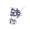
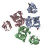
| Entry | Database: PDB / ID: 1sot | ||||||
|---|---|---|---|---|---|---|---|
| Title | Crystal Structure of the DegS stress sensor | ||||||
 Components Components | Protease degS | ||||||
 Keywords Keywords | HYDROLASE / stress response / protein quality control / PDZ / UPR / HtrA | ||||||
| Function / homology |  Function and homology information Function and homology information: / peptidase Do / cellular response to misfolded protein / membrane => GO:0016020 / serine-type peptidase activity / peptidase activity / outer membrane-bounded periplasmic space / serine-type endopeptidase activity / proteolysis / identical protein binding / plasma membrane Similarity search - Function | ||||||
| Biological species |  | ||||||
| Method |  X-RAY DIFFRACTION / SYNCHROTRON / MOLECULAR REPLACEMENT / Resolution: 2.3 Å X-RAY DIFFRACTION / SYNCHROTRON / MOLECULAR REPLACEMENT / Resolution: 2.3 Å | ||||||
 Authors Authors | Wilken, C. / Kitzing, K. / Kurzbauer, R. / Ehrmann, M. / Clausen, T. | ||||||
 Citation Citation | Journal: Cell(Cambridge,Mass.) / Year: 2004 Title: Crystal structure of the DegS stress sensor: How a PDZ domain recognizes misfolded protein and activates a protease Authors: Wilken, C. / Kitzing, K. / Kurzbauer, R. / Ehrmann, M. / Clausen, T. #1: Journal: Cell(Cambridge,Mass.) / Year: 2003Title: OMP peptide signals initiate the envelope stress response by activating DegS protease via relief of inhibition mediated by its PDZ domain Authors: Walsh, N.P. / Alba, B.M. / Bose, B. / Gross, C.A. / Sauer, R.T. | ||||||
| History |
|
- Structure visualization
| Structure viewer | Molecule: MolmilJmol/JSmol |
|---|
- Downloads & links
-Download
| PDBx/mmCIF format | 1sot.cif.gz | 177.6 KB | Display | PDBx/mmCIF format |
|---|---|---|---|---|
| PDB format | pdb1sot.ent.gz | 140.8 KB | Display | PDB format |
| PDBx/mmJSON format | 1sot.json.gz | Tree view | PDBx/mmJSON format | |
| Others |  Other downloads Other downloads |
-Validation report
| Arichive directory | https://data.pdbj.org/pub/pdb/validation_reports/so/1sotftp://data.pdbj.org/pub/pdb/validation_reports/so/1sot | HTTPS FTP |
|---|
-Related structure data
| Related structure data | 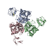 1sozC 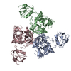 1vcwC  1ky9S S: Starting model for refinement C: citing same article ( |
|---|---|
| Similar structure data |


-Links
 PDBj
PDBj
- Assembly
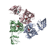
| Deposited unit | 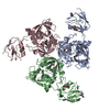
| ||||||||
|---|---|---|---|---|---|---|---|---|---|
| 1 |
| ||||||||
| Unit cell |
|
-Components
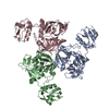
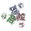
| #1: Protein | Mass: 34353.898 Da / Num. of mol.: 3 / Fragment: protease plus PDZ domain Source method: isolated from a genetically manipulated source Source: (gene. exp.) References: UniProt: P31137, UniProt: P0AEE3*PLUS, Hydrolases; Acting on peptide bonds (peptidases); Serine endopeptidases #2: Water | ChemComp-HOH / |  Mass: 18.015 Da / Num. of mol.: 330 / Source method: isolated from a natural source / Formula: H2O Mass: 18.015 Da / Num. of mol.: 330 / Source method: isolated from a natural source / Formula: H2OHas protein modification | Y | |
|---|
-Experimental details
-Experiment
| Experiment | Method: X-RAY DIFFRACTION / Number of used crystals: 1 |
|---|
- Sample preparation
| Crystal | Density Matthews: 3.51 Å3/Da / Density % sol: 64.63 % |
|---|---|
| Crystal grow | Temperature: 292 K / Method: vapor diffusion, sitting drop / pH: 7.5 Details: PEG 6000, MPD, magnesium chloride, HEPES, pH 7.5, VAPOR DIFFUSION, SITTING DROP, temperature 292.K |
-Data collection
| Diffraction | Mean temperature: 100 K |
|---|---|
| Diffraction source | Source: SYNCHROTRON / Site: ESRF  / Beamline: ID14-4 / Wavelength: 0.9393 Å / Beamline: ID14-4 / Wavelength: 0.9393 Å |
| Detector | Type: ADSC QUANTUM 4 / Detector: CCD / Date: Jun 12, 2003 |
| Radiation | Protocol: SINGLE WAVELENGTH / Monochromatic (M) / Laue (L): M / Scattering type: x-ray |
| Radiation wavelength | Wavelength: 0.9393 Å / Relative weight: 1 |
| Reflection | Resolution: 2.3→15 Å / Num. all: 51588 / Num. obs: 51588 / % possible obs: 96.5 % / Observed criterion σ(F): 0 / Observed criterion σ(I): 0 / Rsym value: 0.049 / Net I/σ(I): 24.1 |
| Reflection shell | Resolution: 2.3→2.34 Å / Redundancy: 1.93 % / Mean I/σ(I) obs: 2.4 / Rsym value: 0.205 / % possible all: 99.5 |
- Processing
| Software |
| ||||||||||||||||||||
|---|---|---|---|---|---|---|---|---|---|---|---|---|---|---|---|---|---|---|---|---|---|
| Refinement | Method to determine structure: MOLECULAR REPLACEMENT Starting model: PDB entry 1KY9 Resolution: 2.3→15 Å / σ(F): 0 / σ(I): 0 / Stereochemistry target values: Engh & Huber
| ||||||||||||||||||||
| Refinement step | Cycle: LAST / Resolution: 2.3→15 Å
| ||||||||||||||||||||
| Refine LS restraints |
|
