 Movie
Movie Controller

[English] 日本語
 Yorodumi
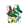
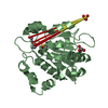
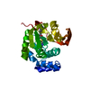
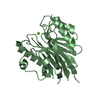
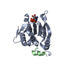
- PDB-1l1j: Crystal structure of the protease domain of an ATP-independent he... -
+ Open data

- Basic information
| Entry | Database: PDB / ID: 1l1j | ||||||
|---|---|---|---|---|---|---|---|
| Title | Crystal structure of the protease domain of an ATP-independent heat shock protease HtrA | ||||||
 Components Components | heat shock protease HtrA | ||||||
 Keywords Keywords | HYDROLASE / serine proteinase | ||||||
| Function / homology |  Function and homology information Function and homology information | ||||||
| Biological species |   Thermotoga maritima (bacteria) Thermotoga maritima (bacteria) | ||||||
| Method |  X-RAY DIFFRACTION / Resolution: 2.8 Å X-RAY DIFFRACTION / Resolution: 2.8 Å | ||||||
 Authors Authors | Kim, D.Y. / Kim, D.R. / Ha, S.C. / Lokanath, N.K. / Hwang, H.Y. / Kim, K.K. | ||||||
 Citation Citation | Journal: J.BIOL.CHEM. / Year: 2003 Title: Crystal Structure of the Protease Domain of a Heat-shock Protein HtrA from Thermotoga maritima Authors: Kim, D.Y. / Kim, D.R. / Ha, S.C. / Lokanath, N.K. / Lee, C.J. / Hwang, H.Y. / Kim, K.K. | ||||||
| History |
|
- Structure visualization
| Structure viewer | Molecule: MolmilJmol/JSmol |
|---|
- Downloads & links
-Download
| PDBx/mmCIF format | 1l1j.cif.gz | 96.7 KB | Display | PDBx/mmCIF format |
|---|---|---|---|---|
| PDB format | pdb1l1j.ent.gz | 75.9 KB | Display | PDB format |
| PDBx/mmJSON format | 1l1j.json.gz | Tree view | PDBx/mmJSON format | |
| Others |  Other downloads Other downloads |
-Validation report
| Arichive directory | https://data.pdbj.org/pub/pdb/validation_reports/l1/1l1jftp://data.pdbj.org/pub/pdb/validation_reports/l1/1l1j | HTTPS FTP |
|---|
-Related structure data
| Similar structure data |
|---|


-Links
 PDBj
PDBj
- Assembly
| Deposited unit | 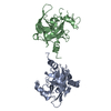
| ||||||||
|---|---|---|---|---|---|---|---|---|---|
| 1 | 
| ||||||||
| 2 | 
| ||||||||
| Unit cell |
|
-Components
| #1: Protein | Mass: 25931.455 Da / Num. of mol.: 2 / Fragment: protease domain Source method: isolated from a genetically manipulated source Source: (gene. exp.) Thermotoga maritima (bacteria) / Plasmid: pET-22b / Production host: References: UniProt: Q9WZ41, Hydrolases; Acting on peptide bonds (peptidases); Serine endopeptidases #2: Water | ChemComp-HOH / |  Mass: 18.015 Da / Num. of mol.: 58 / Source method: isolated from a natural source / Formula: H2O Mass: 18.015 Da / Num. of mol.: 58 / Source method: isolated from a natural source / Formula: H2O |
|---|
-Experimental details
-Experiment
| Experiment | Method: X-RAY DIFFRACTION |
|---|
- Sample preparation
| Crystal | Density Matthews: 2.81 Å3/Da / Density % sol: 56.28 % | ||||||||||||||||||||||||||||
|---|---|---|---|---|---|---|---|---|---|---|---|---|---|---|---|---|---|---|---|---|---|---|---|---|---|---|---|---|---|
| Crystal grow | Temperature: 295 K / Method: vapor diffusion, hanging drop / pH: 4.4 Details: phosphate-citrate, Li2SO4, PEG1000, pH 4.4, VAPOR DIFFUSION, HANGING DROP, temperature 295K | ||||||||||||||||||||||||||||
| Crystal grow | *PLUS Temperature: 22 ℃ | ||||||||||||||||||||||||||||
| Components of the solutions | *PLUS
|
-Data collection
| Radiation | Protocol: SINGLE WAVELENGTH / Monochromatic (M) / Laue (L): M / Scattering type: x-ray |
|---|---|
| Radiation wavelength | Relative weight: 1 |
| Reflection | *PLUS Highest resolution: 2.8 Å / Lowest resolution: 20 Å / Num. obs: 13843 / % possible obs: 95.1 % / Redundancy: 2.43 % / Rmerge(I) obs: 0.066 |
| Reflection shell | *PLUS Highest resolution: 2.8 Å / Lowest resolution: 2.9 Å / % possible obs: 97.2 % / Num. unique obs: 1376 / Rmerge(I) obs: 0.27 |
- Processing
| Software | Name: CNS / Version: 1 / Classification: refinement | ||||||||||||
|---|---|---|---|---|---|---|---|---|---|---|---|---|---|
| Refinement | Resolution: 2.8→20 Å
| ||||||||||||
| Refinement step | Cycle: LAST / Resolution: 2.8→20 Å
| ||||||||||||
| Refinement | *PLUS Num. reflection all: 13621 / Rfactor Rfree: 0.284 / Rfactor Rwork: 0.222 | ||||||||||||
| Solvent computation | *PLUS | ||||||||||||
| Displacement parameters | *PLUS | ||||||||||||
| Refine LS restraints | *PLUS
|
