 Movie
Movie Controller

[English] 日本語
 Yorodumi
- PDB-2yxq: The plug domain of the SecY protein stablizes the closed state of... -
+ Open data

- Basic information
| Entry | Database: PDB / ID: 2yxq | ||||||
|---|---|---|---|---|---|---|---|
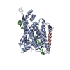
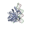
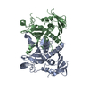
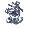
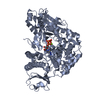
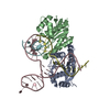
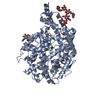
| Title | The plug domain of the SecY protein stablizes the closed state of the translocation channel and maintains a membrane seal | ||||||
 Components Components |
| ||||||
 Keywords Keywords | PROTEIN TRANSPORT / protein translocation / signal peptide / membrane protein / protein secretion / prl mutation | ||||||
| Function / homology |  Function and homology information Function and homology informationintracellular protein transmembrane transport / SRP-dependent cotranslational protein targeting to membrane, translocation / signal sequence receptor activity / protein secretion / transmembrane protein transporter activity / protein targeting / protein transport / plasma membrane Similarity search - Function | ||||||
| Biological species |   Methanocaldococcus jannaschii (archaea) Methanocaldococcus jannaschii (archaea) | ||||||
| Method |  X-RAY DIFFRACTION / SYNCHROTRON / MOLECULAR REPLACEMENT / Resolution: 3.5 Å X-RAY DIFFRACTION / SYNCHROTRON / MOLECULAR REPLACEMENT / Resolution: 3.5 Å | ||||||
 Authors Authors | Li, W. / Schulman, S. | ||||||
 Citation Citation | Journal: Mol.Cell / Year: 2007 Title: The plug domain of the SecY protein stabilizes the closed state of the translocation channel and maintains a membrane seal Authors: Li, W. / Schulman, S. / Boyd, D. / Erlandson, K. / Beckwith, J. / Rapoport, T.A. | ||||||
| History |
| ||||||
| Remark 999 | SEQUENCE deletion of 60-65 and substitution with one Gly |
- Structure visualization
| Structure viewer | Molecule: MolmilJmol/JSmol |
|---|
- Downloads & links
-Download
| PDBx/mmCIF format | 2yxq.cif.gz | 106.6 KB | Display | PDBx/mmCIF format |
|---|---|---|---|---|
| PDB format | pdb2yxq.ent.gz | 82 KB | Display | PDB format |
| PDBx/mmJSON format | 2yxq.json.gz | Tree view | PDBx/mmJSON format | |
| Others |  Other downloads Other downloads |
-Validation report
| Arichive directory | https://data.pdbj.org/pub/pdb/validation_reports/yx/2yxqftp://data.pdbj.org/pub/pdb/validation_reports/yx/2yxq | HTTPS FTP |
|---|
-Related structure data
| Related structure data | 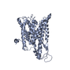 2yxrC 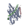 1rhzS S: Starting model for refinement C: citing same article ( |
|---|---|
| Similar structure data |


-Links
 PDBj
PDBj
- Assembly
| Deposited unit | 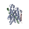
| ||||||||
|---|---|---|---|---|---|---|---|---|---|
| 1 | 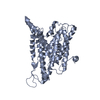
| ||||||||
| 2 | 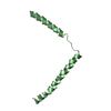
| ||||||||
| 3 | 
| ||||||||
| 4 |
| ||||||||
| Unit cell |
| ||||||||
| Details | The biological assembly is a monomer in the asymmetric unit |
-Components
| #1: Protein | Mass: 46938.301 Da / Num. of mol.: 1 Source method: isolated from a genetically manipulated source Source: (gene. exp.) Methanocaldococcus jannaschii (archaea)Gene: secY / Plasmid: pBAD / Production host:  |
|---|---|
| #2: Protein | Mass: 8451.144 Da / Num. of mol.: 1 Source method: isolated from a genetically manipulated source Source: (gene. exp.) Methanocaldococcus jannaschii (archaea)Gene: secE / Plasmid: pBAD / Production host: |
| #3: Protein | Mass: 5967.010 Da / Num. of mol.: 1 Source method: isolated from a genetically manipulated source Source: (gene. exp.) Methanocaldococcus jannaschii (archaea)Gene: secG / Plasmid: pBAD / Production host: |
-Experimental details
-Experiment
| Experiment | Method: X-RAY DIFFRACTION / Number of used crystals: 1 |
|---|
- Sample preparation
| Crystal | Density Matthews: 4.82 Å3/Da / Density % sol: 74.51 % |
|---|---|
| Crystal grow | Temperature: 277 K / Method: vapor diffusion, hanging drop / pH: 9 Details: 40-55% PEG400, 50mM Glycine-HCl , pH9, VAPOR DIFFUSION, HANGING DROP, temperature 277K |
-Data collection
| Diffraction | Mean temperature: 200 K |
|---|---|
| Diffraction source | Source: SYNCHROTRON / Site: APS  / Beamline: 19-ID / Wavelength: 0.97921 Å / Beamline: 19-ID / Wavelength: 0.97921 Å |
| Detector | Type: ADSC QUANTUM 315 / Detector: CCD / Date: Dec 11, 2006 |
| Radiation | Monochromator: Si 111 / Protocol: SINGLE WAVELENGTH / Monochromatic (M) / Laue (L): M / Scattering type: x-ray |
| Radiation wavelength | Wavelength: 0.97921 Å / Relative weight: 1 |
| Reflection | Resolution: 3.5→50 Å / Num. all: 14806 / Num. obs: 14678 / % possible obs: 98.6 % / Observed criterion σ(F): 1.5 / Observed criterion σ(I): 2.3 / Redundancy: 12 % / Biso Wilson estimate: 118.6 Å2 / Rmerge(I) obs: 0.087 / Rsym value: 0.087 / Net I/σ(I): 21.9 |
| Reflection shell | Resolution: 3.5→3.63 Å / Redundancy: 12 % / Rmerge(I) obs: 0.787 / Mean I/σ(I) obs: 2.3 / Num. unique all: 1419 / Rsym value: 0.787 / % possible all: 97.5 |
- Processing
| Software |
| ||||||||||||||||||||||||||||||||||||||||||
|---|---|---|---|---|---|---|---|---|---|---|---|---|---|---|---|---|---|---|---|---|---|---|---|---|---|---|---|---|---|---|---|---|---|---|---|---|---|---|---|---|---|---|---|
| Refinement | Method to determine structure: MOLECULAR REPLACEMENT Starting model: 1RHZ Resolution: 3.5→50 Å / Isotropic thermal model: Isotropic / Cross valid method: THROUGHOUT / σ(F): 1.5 / σ(I): 2.3 / Stereochemistry target values: Engh & Huber
| ||||||||||||||||||||||||||||||||||||||||||
| Displacement parameters | Biso mean: -0.367 Å2
| ||||||||||||||||||||||||||||||||||||||||||
| Refinement step | Cycle: LAST / Resolution: 3.5→50 Å
| ||||||||||||||||||||||||||||||||||||||||||
| LS refinement shell |
|
