Mass: 18.015 Da / Num. of mol.: 277 / Source method: isolated from a natural source / Formula: H2O
Sequence details
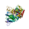
THE FIRST FIVE RESIDUES )IN CHAIN A (MNNRN) AND SEVEN IN CHAIN B (MNNRNHD) RESIDUES ARE DISORDERED. ...THE FIRST FIVE RESIDUES )IN CHAIN A (MNNRN) AND SEVEN IN CHAIN B (MNNRNHD) RESIDUES ARE DISORDERED. RESIDUES 99 (Q) AND 572 - 577 (AEADRQ) IN CHAIN A ARE DISORDERED. IN CHAIN B RESIDUES 98 AND 99 (AQ)ARE DISORDERED. IN BOTH CHAINS THE TERMINAL REDIDUES 587-620 ( GHAVQHGSEVQHDERRHGDVRHEEARHGEVQHG) ARE DISORDERED
-
Experimental details
-
Experiment
Experiment
Method: X-RAY DIFFRACTION / Number of used crystals: 1
-
Sample preparation
Crystal
Density Matthews: 2.13 Å3/Da / Density % sol: 42 % / Description: NONE
Crystal grow
Temperature: 298 K / Method: vapor diffusion, hanging drop / pH: 7.8 Details: 0.1 M TRIS-HCL PH 7.8, 26% (W/V) PEG8000, 300 MM L-SERINE. 5MM ATP (DISSOLVED IN WATER) WAS PRE-INCUBATED FOR 10 MIN (RT) WITH 6 MG/ML ACSD. PROTEINULLTP PRECIPITATE WAS REMOVED BY ...Details: 0.1 M TRIS-HCL PH 7.8, 26% (W/V) PEG8000, 300 MM L-SERINE. 5MM ATP (DISSOLVED IN WATER) WAS PRE-INCUBATED FOR 10 MIN (RT) WITH 6 MG/ML ACSD. PROTEINULLTP PRECIPITATE WAS REMOVED BY CENTRIFUGATION. 1 UL OF SUPERNATANT AND EQUAL AMOUNT OF PRECIPITANT WAS USED IN HANGING DROP CRYSTALLIZATION (298 K).
Monochromator: DIAMOND (111), GE(220) / Protocol: SINGLE WAVELENGTH / Monochromatic (M) / Laue (L): M / Scattering type: x-ray
Radiation wavelength
Wavelength: 0.933 Å / Relative weight: 1
Reflection
Resolution: 2.2→81.1 Å / Num. obs: 61355 / % possible obs: 99.9 % / Observed criterion σ(I): 2 / Redundancy: 10.1 % / Rmerge(I) obs: 0.11 / Net I/σ(I): 15.7
Reflection shell
Resolution: 2.2→2.95 Å / Redundancy: 7.3 % / Rmerge(I) obs: 0.38 / Mean I/σ(I) obs: 5.9 / % possible all: 99.9
-
Processing
Software
Name
Version
Classification
REFMAC
5.2.0019
refinement
XDS
datareduction
XSCALA
datascaling
PHASER
phasing
Refinement
Method to determine structure: MOLECULAR REPLACEMENT Starting model: APO STRUCTURE OF ACSD Resolution: 2.2→81.11 Å / Cor.coef. Fo:Fc: 0.936 / Cor.coef. Fo:Fc free: 0.884 / SU B: 20.286 / SU ML: 0.243 / Cross valid method: THROUGHOUT / ESU R: 0.336 / ESU R Free: 0.252 / Stereochemistry target values: MAXIMUM LIKELIHOOD / Details: HYDROGENS HAVE BEEN ADDED IN THE RIDING POSITIONS.
Rfactor
Num. reflection
% reflection
Selection details
Rfree
0.277
3096
5 %
RANDOM
Rwork
0.216
-
-
-
obs
0.219
58259
99.9 %
-
Solvent computation
Ion probe radii: 0.8 Å / Shrinkage radii: 0.8 Å / VDW probe radii: 1.2 Å / Solvent model: BABINET MODEL WITH MASK
 Movie
Movie Controller

 Yorodumi
 Open data
 Basic information
 Components
 Keywords
 Function and homology information
 ERWINIA CHRYSANTHEMI (bacteria)
 X-RAY DIFFRACTION /
  Authors
 Citation
 Structure visualization
 Downloads & links
 Other downloads


 PDBj
PDBj Assembly
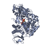
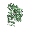

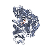
 Mass: 507.181 Da / Num. of mol.: 2 / Source method: obtained synthetically / Formula: C10H16N5O13P3 / Comment: ATP, energy-carrying molecule*YM
 Mass: 24.305 Da / Num. of mol.: 2 / Source method: obtained synthetically / Formula: Mg
 Type: L-peptide linking / Mass: 105.093 Da / Num. of mol.: 1 / Source method: obtained synthetically / Formula: C3H7NO3
 Mass: 18.015 Da / Num. of mol.: 277 / Source method: isolated from a natural source / Formula: H2O
 Sample preparation
 / Beamline: ID14-2 / Wavelength: 0.933
  Processing