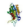
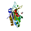
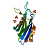
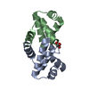
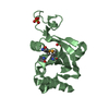
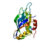
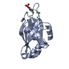
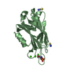
Entry Database : PDB / ID : 2yoaTitle Synaptotagmin-1 C2B domain with phosphoserine SYNAPTOTAGMIN-1 Keywords Function / homology Function Domain/homology Component
/ / / / / / / / / / / / / / / / / / / / / / / / / / / / / / / / / / / / / / / / / / / / / / / / / / / / / / / / / / / / / / / / / / / / / / / / / / / / / / / / / / / / / / / / / / / / / / / / / / / / / / / / Biological species RATTUS NORVEGICUS (Norway rat)Method / / / Resolution : 1.5 Å Authors Honigmann, A. / van den Bogaart, G. / Iraheta, E. / Risselada, H.J. / Milovanovic, D. / Mueller, V. / Muellar, S. / Diederichsen, U. / Fasshauer, D. / Grubmuller, H. ...Honigmann, A. / van den Bogaart, G. / Iraheta, E. / Risselada, H.J. / Milovanovic, D. / Mueller, V. / Muellar, S. / Diederichsen, U. / Fasshauer, D. / Grubmuller, H. / Hell, S.W. / Eggeling, C. / Kuhnel, K. / Jahn, R. Journal : Nat.Struct.Mol.Biol. / Year : 2013Title : Phosphatidylinositol 4,5-Bisphosphate Clusters Act as Molecular Beacons for Vesicle RecruitmentAuthors: Honigmann, A. / Van Den Bogaart, G. / Iraheta, E. / Risselada, H.J. / Milovanovic, D. / Mueller, V. / Muellar, S. / Diederichsen, U. / Fasshauer, D. / Grubmuller, H. / Hell, S.W. / Eggeling, ... Authors : Honigmann, A. / Van Den Bogaart, G. / Iraheta, E. / Risselada, H.J. / Milovanovic, D. / Mueller, V. / Muellar, S. / Diederichsen, U. / Fasshauer, D. / Grubmuller, H. / Hell, S.W. / Eggeling, C. / Kuhnel, K. / Jahn, R. History Deposition Oct 22, 2012 Deposition site / Processing site Revision 1.0 Mar 20, 2013 Provider / Type Revision 1.1 May 15, 2013 Group Revision 1.2 May 29, 2013 Group Revision 1.3 Jun 19, 2013 Group Revision 1.4 Aug 23, 2017 Group / Category / Item Revision 1.5 Dec 20, 2023 Group Data collection / Database references ... Data collection / Database references / Derived calculations / Other / Refinement description Category chem_comp_atom / chem_comp_bond ... chem_comp_atom / chem_comp_bond / database_2 / pdbx_database_status / pdbx_initial_refinement_model / pdbx_struct_conn_angle / struct_conn / struct_site Item _database_2.pdbx_DOI / _database_2.pdbx_database_accession ... _database_2.pdbx_DOI / _database_2.pdbx_database_accession / _pdbx_database_status.status_code_sf / _pdbx_struct_conn_angle.ptnr1_auth_comp_id / _pdbx_struct_conn_angle.ptnr1_auth_seq_id / _pdbx_struct_conn_angle.ptnr1_label_asym_id / _pdbx_struct_conn_angle.ptnr1_label_atom_id / _pdbx_struct_conn_angle.ptnr1_label_comp_id / _pdbx_struct_conn_angle.ptnr1_label_seq_id / _pdbx_struct_conn_angle.ptnr2_auth_seq_id / _pdbx_struct_conn_angle.ptnr2_label_asym_id / _pdbx_struct_conn_angle.ptnr3_auth_comp_id / _pdbx_struct_conn_angle.ptnr3_auth_seq_id / _pdbx_struct_conn_angle.ptnr3_label_asym_id / _pdbx_struct_conn_angle.ptnr3_label_atom_id / _pdbx_struct_conn_angle.ptnr3_label_comp_id / _pdbx_struct_conn_angle.ptnr3_label_seq_id / _pdbx_struct_conn_angle.value / _struct_conn.pdbx_dist_value / _struct_conn.ptnr1_auth_comp_id / _struct_conn.ptnr1_auth_seq_id / _struct_conn.ptnr1_label_asym_id / _struct_conn.ptnr1_label_atom_id / _struct_conn.ptnr1_label_comp_id / _struct_conn.ptnr1_label_seq_id / _struct_conn.ptnr2_auth_comp_id / _struct_conn.ptnr2_auth_seq_id / _struct_conn.ptnr2_label_asym_id / _struct_conn.ptnr2_label_atom_id / _struct_conn.ptnr2_label_comp_id / _struct_conn.ptnr2_label_seq_id / _struct_site.pdbx_auth_asym_id / _struct_site.pdbx_auth_comp_id / _struct_site.pdbx_auth_seq_id
Show all Show less
 Movie
Movie Controller


 Open data
 Basic information
 Components
 Keywords
 Function and homology information
 X-RAY DIFFRACTION /
  Authors
 Citation
 Structure visualization
 Downloads & links
 Other downloads


 PDBj
PDBj


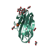
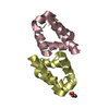
 Assembly
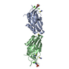


 Mass: 40.078 Da / Num. of mol.: 7 / Source method: obtained synthetically / Formula: Ca
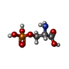
 Type: L-peptide linking / Mass: 185.072 Da / Num. of mol.: 2 / Source method: obtained synthetically / Formula: C3H8NO6P
 Mass: 58.082 Da / Num. of mol.: 5 / Source method: obtained synthetically / Formula: CNS
 Mass: 18.015 Da / Num. of mol.: 256 / Source method: isolated from a natural source / Formula: H2O
 Sample preparation
 / Beamline: X10SA / Wavelength: 0.99998
  Processing