 Movie
Movie Controller


+ Open data

- Basic information
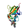
| Entry | Database: PDB / ID: 1uow | ||||||
|---|---|---|---|---|---|---|---|
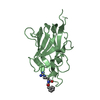
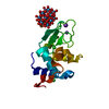
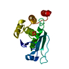
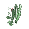
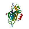
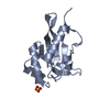
| Title | Calcium binding domain C2B | ||||||
 Components Components | SYNAPTOTAGMIN I | ||||||
 Keywords Keywords | GLYCOPROTEIN / LIPOPROTEIN / TRANSMEMBRANE | ||||||
| Function / homology |  Function and homology information Function and homology informationsynchronous neurotransmitter secretion / regulation of vesicle fusion / spontaneous neurotransmitter secretion / fast, calcium ion-dependent exocytosis of neurotransmitter / syntaxin-3 binding / regulation of regulated secretory pathway / regulation of calcium-dependent activation of synaptic vesicle fusion / calcium-dependent activation of synaptic vesicle fusion / chromaffin granule membrane / : ...synchronous neurotransmitter secretion / regulation of vesicle fusion / spontaneous neurotransmitter secretion / fast, calcium ion-dependent exocytosis of neurotransmitter / syntaxin-3 binding / regulation of regulated secretory pathway / regulation of calcium-dependent activation of synaptic vesicle fusion / calcium-dependent activation of synaptic vesicle fusion / chromaffin granule membrane / : / positive regulation of calcium ion-dependent exocytosis of neurotransmitter / calcium activated phospholipid scrambling / calcium ion-regulated exocytosis of neurotransmitter / Glutamate Neurotransmitter Release Cycle / Norepinephrine Neurotransmitter Release Cycle / Acetylcholine Neurotransmitter Release Cycle / Serotonin Neurotransmitter Release Cycle / GABA synthesis, release, reuptake and degradation / regulation of calcium ion-dependent exocytosis / Dopamine Neurotransmitter Release Cycle / calcium ion sensor activity / synaptic vesicle recycling / calcium-ion regulated exocytosis / exocytic vesicle / positive regulation of calcium ion-dependent exocytosis / protein heterooligomerization / vesicle organization / : / positive regulation of vesicle fusion / positive regulation of dendrite extension / Cargo recognition for clathrin-mediated endocytosis / Clathrin-mediated endocytosis / vesicle fusion / calcium-dependent phospholipid binding / positive regulation of dopamine secretion / negative regulation of neurotransmitter secretion / dense core granule / membraneless organelle assembly / xenobiotic transmembrane transport / neurotransmitter secretion / syntaxin binding / syntaxin-1 binding / presynaptic active zone / inhibitory postsynaptic potential / low-density lipoprotein particle receptor binding / phosphatidylserine binding / clathrin binding / regulation of synaptic vesicle exocytosis / neuron projection terminus / regulation of dopamine secretion / protein insertion into membrane / regulation of endocytosis / synaptic vesicle exocytosis / detection of calcium ion / postsynaptic cytosol / positive regulation of synaptic transmission / regulation of synaptic transmission, glutamatergic / presynaptic cytosol / vesicle-mediated transport / phosphatidylinositol-4,5-bisphosphate binding / excitatory synapse / secretory granule / cellular response to calcium ion / hippocampal mossy fiber to CA3 synapse / SNARE binding / molecular condensate scaffold activity / phospholipid binding / response to calcium ion / calcium-dependent protein binding / terminal bouton / synaptic vesicle / synaptic vesicle membrane / presynaptic membrane / cell differentiation / calmodulin binding / postsynaptic membrane / neuron projection / postsynaptic density / protein heterodimerization activity / axon / calcium ion binding / lipid binding / glutamatergic synapse / Golgi apparatus / identical protein binding / plasma membrane / cytoplasm Similarity search - Function | ||||||
| Biological species |  | ||||||
| Method |  X-RAY DIFFRACTION / SYNCHROTRON / MOLECULAR REPLACEMENT / Resolution: 1.04 Å X-RAY DIFFRACTION / SYNCHROTRON / MOLECULAR REPLACEMENT / Resolution: 1.04 Å | ||||||
 Authors Authors | Cheng, Y. / Sequeira, S.M. / Sollner, T.H. / Patel, D.J. | ||||||
 Citation Citation | Journal: Protein Sci. / Year: 2004 Title: Crystallographic Identification of Ca2+ and Sr2+ Coordination Sites in Synaptotagmin I C2B Domain Authors: Cheng, Y. / Sequeira, S.M. / Malinina, L. / Tereshko, V. / Sollner, T.H. / Patel, D.J. | ||||||
| History |
|
- Structure visualization
| Structure viewer | Molecule: MolmilJmol/JSmol |
|---|
- Downloads & links
-Download
| PDBx/mmCIF format | 1uow.cif.gz | 85 KB | Display | PDBx/mmCIF format |
|---|---|---|---|---|
| PDB format | pdb1uow.ent.gz | 62.5 KB | Display | PDB format |
| PDBx/mmJSON format | 1uow.json.gz | Tree view | PDBx/mmJSON format | |
| Others |  Other downloads Other downloads |
-Validation report
| Arichive directory | https://data.pdbj.org/pub/pdb/validation_reports/uo/1uowftp://data.pdbj.org/pub/pdb/validation_reports/uo/1uow | HTTPS FTP |
|---|
-Related structure data
| Related structure data | 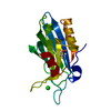 1tjmC 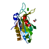 1tjxC  1uovC  1k5wS S: Starting model for refinement C: citing same article ( |
|---|---|
| Similar structure data |


-Links
 PDBj
PDBj


- Assembly
| Deposited unit | 
| ||||||||
|---|---|---|---|---|---|---|---|---|---|
| 1 |
| ||||||||
| Unit cell |
|
-Components
| #1: Protein | Mass: 17822.727 Da / Num. of mol.: 1 / Fragment: C2B DOMAIN, RESIDUES 271-421 Source method: isolated from a genetically manipulated source Details: 2 CALCIUM IONS BOUND, THIS STRUCTURE IS AT THE SAME RESOLUTION AS PDB ENTRY 1TJX, BUT THE LATTER CONTAINS DEPOSITED PROTONS Source: (gene. exp.)  | ||||||||
|---|---|---|---|---|---|---|---|---|---|
| #2: Chemical |   Mass: 40.078 Da / Num. of mol.: 2 / Source method: obtained synthetically / Formula: Ca Mass: 40.078 Da / Num. of mol.: 2 / Source method: obtained synthetically / Formula: Ca#3: Chemical | ChemComp-GOL / |   Mass: 92.094 Da / Num. of mol.: 1 / Source method: obtained synthetically / Formula: C3H8O3 Mass: 92.094 Da / Num. of mol.: 1 / Source method: obtained synthetically / Formula: C3H8O3#4: Chemical | ChemComp-ACT / |   Mass: 59.044 Da / Num. of mol.: 1 / Source method: obtained synthetically / Formula: C2H3O2 Mass: 59.044 Da / Num. of mol.: 1 / Source method: obtained synthetically / Formula: C2H3O2#5: Water | ChemComp-HOH / |  Mass: 18.015 Da / Num. of mol.: 184 / Source method: isolated from a natural source / Formula: H2O Mass: 18.015 Da / Num. of mol.: 184 / Source method: isolated from a natural source / Formula: H2OCompound details | COULD PLAY A REGULATORY ROLE IN THE MEMBRANE INTERACTIONS DURING TRAFFICKING OF SYNAPTIC VESICLES ...COULD PLAY A REGULATORY | |
-Experimental details
-Experiment
| Experiment | Method: X-RAY DIFFRACTION / Number of used crystals: 1 |
|---|
- Sample preparation
| Crystal | Density Matthews: 2.5 Å3/Da / Density % sol: 50.73 % |
|---|---|
| Crystal grow | pH: 5 / Details: pH 5.00 |
-Data collection
| Diffraction | Mean temperature: 100 K |
|---|---|
| Diffraction source | Source: SYNCHROTRON / Site: APS  / Beamline: 14-BM-C / Wavelength: 0.9 / Beamline: 14-BM-C / Wavelength: 0.9 |
| Detector | Date: May 15, 2003 |
| Radiation | Protocol: SINGLE WAVELENGTH / Monochromatic (M) / Laue (L): M / Scattering type: x-ray |
| Radiation wavelength | Wavelength: 0.9 Å / Relative weight: 1 |
| Reflection | Resolution: 1.04→20 Å / Num. obs: 4126 / % possible obs: 99 % / Redundancy: 5.6 % / Rmerge(I) obs: 0.103 / Net I/σ(I): 19.34 |
| Reflection shell | Resolution: 1.04→1.06 Å / Rmerge(I) obs: 0.416 / Mean I/σ(I) obs: 1.62 / % possible all: 96.6 |
- Processing
| Software |
| ||||||||||||||||||||
|---|---|---|---|---|---|---|---|---|---|---|---|---|---|---|---|---|---|---|---|---|---|
| Refinement | Method to determine structure: MOLECULAR REPLACEMENT Starting model: PDB ENTRY 1K5W Resolution: 1.04→20 Å / SU B: 0.332 / SU ML: 0.017 / Cross valid method: THROUGHOUT / ESU R: 0.026 / ESU R Free: 0.026
| ||||||||||||||||||||
| Displacement parameters | Biso mean: 12.317 Å2
| ||||||||||||||||||||
| Refinement step | Cycle: LAST / Resolution: 1.04→20 Å
|
