 Movie
Movie Controller

[English] 日本語
 Yorodumi
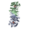
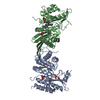
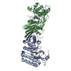
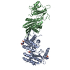
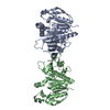
- PDB-2ybx: Crystal Structure of Human Phosphatidylinositol-5-phosphate 4-kin... -
+ Open data

- Basic information
| Entry | Database: PDB / ID: 2ybx | ||||||
|---|---|---|---|---|---|---|---|
| Title | Crystal Structure of Human Phosphatidylinositol-5-phosphate 4-kinase type-2 alpha | ||||||
 Components Components | PHOSPHATIDYLINOSITOL-5-PHOSPHATE 4-KINASE TYPE-2 ALPHA | ||||||
 Keywords Keywords | TRANSFERASE / KINASE / SIGNALLING | ||||||
| Function / homology |  Function and homology information Function and homology informationvesicle-mediated cholesterol transport / 1-phosphatidylinositol-5-phosphate 4-kinase / 1-phosphatidylinositol-5-phosphate 4-kinase activity / 1-phosphatidyl-1D-myo-inositol 4,5-bisphosphate biosynthetic process / Synthesis of PIPs in the nucleus / 1-phosphatidylinositol-4-phosphate 5-kinase activity / autophagosome-lysosome fusion / positive regulation of autophagosome assembly / megakaryocyte development / phosphatidylinositol phosphate biosynthetic process ...vesicle-mediated cholesterol transport / 1-phosphatidylinositol-5-phosphate 4-kinase / 1-phosphatidylinositol-5-phosphate 4-kinase activity / 1-phosphatidyl-1D-myo-inositol 4,5-bisphosphate biosynthetic process / Synthesis of PIPs in the nucleus / 1-phosphatidylinositol-4-phosphate 5-kinase activity / autophagosome-lysosome fusion / positive regulation of autophagosome assembly / megakaryocyte development / phosphatidylinositol phosphate biosynthetic process / PI5P Regulates TP53 Acetylation / Synthesis of PIPs at the plasma membrane / photoreceptor outer segment / photoreceptor inner segment / negative regulation of insulin receptor signaling pathway / autophagosome / regulation of autophagy / PI5P, PP2A and IER3 Regulate PI3K/AKT Signaling / lysosome / protein homodimerization activity / nucleoplasm / ATP binding / plasma membrane / cytosol Similarity search - Function | ||||||
| Biological species |  HOMO SAPIENS (human) HOMO SAPIENS (human) | ||||||
| Method |  X-RAY DIFFRACTION / SYNCHROTRON / MOLECULAR REPLACEMENT / Resolution: 2.56 Å X-RAY DIFFRACTION / SYNCHROTRON / MOLECULAR REPLACEMENT / Resolution: 2.56 Å | ||||||
 Authors Authors | Tresaugues, L. / Moche, M. / Arrowsmith, C.H. / Berglund, H. / Bountra, C. / Collins, R. / Edwards, A.M. / Ekblad, T. / Flodin, S. / Graslund, S. ...Tresaugues, L. / Moche, M. / Arrowsmith, C.H. / Berglund, H. / Bountra, C. / Collins, R. / Edwards, A.M. / Ekblad, T. / Flodin, S. / Graslund, S. / Karlberg, T. / Kotenyova, T. / Kouznetsova, E. / Nyman, T. / Persson, C. / Schuler, H. / Siponen, M.I. / Thorsell, A.G. / Wahlberg, E. / Weigelt, J. / Nordlund, P. | ||||||
 Citation Citation | Journal: To be Published Title: Crystal Structure of Human Phosphatidylinositol-5-Phosphate 4-Kinase Type-2 Alpha Authors: Tresaugues, L. / Moche, M. / Arrowsmith, C.H. / Berglund, H. / Bountra, C. / Collins, R. / Edwards, A.M. / Ekblad, T. / Flodin, S. / Graslund, S. / Karlberg, T. / Kotenyova, T. / ...Authors: Tresaugues, L. / Moche, M. / Arrowsmith, C.H. / Berglund, H. / Bountra, C. / Collins, R. / Edwards, A.M. / Ekblad, T. / Flodin, S. / Graslund, S. / Karlberg, T. / Kotenyova, T. / Kouznetsova, E. / Nyman, T. / Persson, C. / Schuler, H. / Siponen, M.I. / Thorsell, A.G. / Wahlberg, E. / Weigelt, J. / Nordlund, P. | ||||||
| History |
|
- Structure visualization
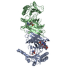
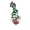
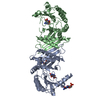
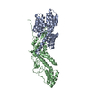
| Structure viewer | Molecule: MolmilJmol/JSmol |
|---|
- Downloads & links
-Download
| PDBx/mmCIF format | 2ybx.cif.gz | 146.4 KB | Display | PDBx/mmCIF format |
|---|---|---|---|---|
| PDB format | pdb2ybx.ent.gz | 112.9 KB | Display | PDB format |
| PDBx/mmJSON format | 2ybx.json.gz | Tree view | PDBx/mmJSON format | |
| Others |  Other downloads Other downloads |
-Validation report
| Arichive directory | https://data.pdbj.org/pub/pdb/validation_reports/yb/2ybxftp://data.pdbj.org/pub/pdb/validation_reports/yb/2ybx | HTTPS FTP |
|---|
-Related structure data
| Related structure data | 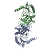 1bo1S S: Starting model for refinement |
|---|---|
| Similar structure data |

-Links
 PDBj
PDBj


- Assembly
| Deposited unit | 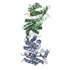
| ||||||||
|---|---|---|---|---|---|---|---|---|---|
| 1 |
| ||||||||
| Unit cell |
| ||||||||
| Noncrystallographic symmetry (NCS) | NCS oper: (Code: given Matrix: (-0.99988, 0.01034, -0.01175), Vector: |
-Components
| #1: Protein | Mass: 45312.984 Da / Num. of mol.: 2 / Fragment: RESIDUES 35-405 Source method: isolated from a genetically manipulated source Details: PHOSPHORYL-ASPARTATE INTERMEDIATE IN POSITION 359 / Source: (gene. exp.) HOMO SAPIENS (human) / Description: MAMMALIAN GENE COLLECTION (MGC) / Plasmid: PNIC28-BSA4 / Production host:  References: UniProt: P48426, 1-phosphatidylinositol-5-phosphate 4-kinase #2: Chemical | ChemComp-PO4 / |   Mass: 94.971 Da / Num. of mol.: 1 / Source method: obtained synthetically / Formula: PO4 Mass: 94.971 Da / Num. of mol.: 1 / Source method: obtained synthetically / Formula: PO4#3: Water | ChemComp-HOH / |  Mass: 18.015 Da / Num. of mol.: 121 / Source method: isolated from a natural source / Formula: H2O Mass: 18.015 Da / Num. of mol.: 121 / Source method: isolated from a natural source / Formula: H2O |
|---|
-Experimental details
-Experiment
| Experiment | Method: X-RAY DIFFRACTION / Number of used crystals: 1 |
|---|
- Sample preparation
| Crystal | Density Matthews: 2.51 Å3/Da / Density % sol: 56.5 % / Description: NONE |
|---|---|
| Crystal grow | pH: 7.5 Details: 0.2 M MAGNESIUM FORMATE, 16% PEG3350, 2 MM BENZYL(1,3,5)TRIPHOSPHATE, pH 7.5 |
-Data collection
| Diffraction | Mean temperature: 100 K |
|---|---|
| Diffraction source | Source: SYNCHROTRON / Site: Diamond  / Beamline: I03 / Wavelength: 0.9792 / Beamline: I03 / Wavelength: 0.9792 |
| Detector | Type: ADSC CCD / Detector: CCD / Date: Jul 8, 2010 |
| Radiation | Protocol: SINGLE WAVELENGTH / Monochromatic (M) / Laue (L): M / Scattering type: x-ray |
| Radiation wavelength | Wavelength: 0.9792 Å / Relative weight: 1 |
| Reflection | Resolution: 2.56→28.47 Å / Num. obs: 28302 / % possible obs: 98.1 % / Observed criterion σ(I): -3 / Redundancy: 3.7 % / Biso Wilson estimate: 55.66 Å2 / Rmerge(I) obs: 0.09 / Net I/σ(I): 7.9 |
| Reflection shell | Resolution: 2.56→2.7 Å / Redundancy: 3.6 % / Rmerge(I) obs: 0.68 / Mean I/σ(I) obs: 1.1 / % possible all: 96.8 |
- Processing
| Software |
| ||||||||||||||||||||||||||||||||||||||||||||||||||||||||||||||||||||||||||||||||||||||||||||||||||||||||||||||||||
|---|---|---|---|---|---|---|---|---|---|---|---|---|---|---|---|---|---|---|---|---|---|---|---|---|---|---|---|---|---|---|---|---|---|---|---|---|---|---|---|---|---|---|---|---|---|---|---|---|---|---|---|---|---|---|---|---|---|---|---|---|---|---|---|---|---|---|---|---|---|---|---|---|---|---|---|---|---|---|---|---|---|---|---|---|---|---|---|---|---|---|---|---|---|---|---|---|---|---|---|---|---|---|---|---|---|---|---|---|---|---|---|---|---|---|---|
| Refinement | Method to determine structure: MOLECULAR REPLACEMENT Starting model: PDB ENTRY 1BO1 Resolution: 2.56→28.37 Å / Cor.coef. Fo:Fc: 0.8973 / Cor.coef. Fo:Fc free: 0.8661 / SU R Cruickshank DPI: 0.428 / Cross valid method: THROUGHOUT / σ(F): 0 / SU R Blow DPI: 0.401 / SU Rfree Blow DPI: 0.259 / SU Rfree Cruickshank DPI: 0.267 Details: IDEAL-DIST CONTACT TERM CONTACT SETUP. ALL ATOMS HAVE CCP4 ATOM TYPE FROM LIBRARY.
| ||||||||||||||||||||||||||||||||||||||||||||||||||||||||||||||||||||||||||||||||||||||||||||||||||||||||||||||||||
| Displacement parameters | Biso mean: 49.11 Å2
| ||||||||||||||||||||||||||||||||||||||||||||||||||||||||||||||||||||||||||||||||||||||||||||||||||||||||||||||||||
| Refine analyze | Luzzati coordinate error obs: 0.314 Å | ||||||||||||||||||||||||||||||||||||||||||||||||||||||||||||||||||||||||||||||||||||||||||||||||||||||||||||||||||
| Refinement step | Cycle: LAST / Resolution: 2.56→28.37 Å
| ||||||||||||||||||||||||||||||||||||||||||||||||||||||||||||||||||||||||||||||||||||||||||||||||||||||||||||||||||
| Refine LS restraints |
| ||||||||||||||||||||||||||||||||||||||||||||||||||||||||||||||||||||||||||||||||||||||||||||||||||||||||||||||||||
| LS refinement shell | Resolution: 2.56→2.66 Å / Total num. of bins used: 14
|
