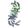
 Movie
Movie Controller


+ Open data

- Basic information
| Entry | Database: PDB / ID: 1bo1 | ||||||
|---|---|---|---|---|---|---|---|
| Title | PHOSPHATIDYLINOSITOL PHOSPHATE KINASE TYPE II BETA | ||||||
 Components Components | PROTEIN (PHOSPHATIDYLINOSITOL PHOSPHATE KINASE IIBETA) | ||||||
 Keywords Keywords | TRANSFERASE / LIPID SIGNALING | ||||||
| Function / homology |  Function and homology information Function and homology information1-phosphatidylinositol-5-phosphate 4-kinase / 1-phosphatidylinositol-5-phosphate 4-kinase activity / 1-phosphatidyl-1D-myo-inositol 4,5-bisphosphate biosynthetic process / Synthesis of PIPs in the nucleus / 1-phosphatidylinositol-4-phosphate 5-kinase activity / autophagosome-lysosome fusion / positive regulation of autophagosome assembly / phosphatidylinositol phosphate biosynthetic process / PI5P Regulates TP53 Acetylation / Synthesis of PIPs at the plasma membrane ...1-phosphatidylinositol-5-phosphate 4-kinase / 1-phosphatidylinositol-5-phosphate 4-kinase activity / 1-phosphatidyl-1D-myo-inositol 4,5-bisphosphate biosynthetic process / Synthesis of PIPs in the nucleus / 1-phosphatidylinositol-4-phosphate 5-kinase activity / autophagosome-lysosome fusion / positive regulation of autophagosome assembly / phosphatidylinositol phosphate biosynthetic process / PI5P Regulates TP53 Acetylation / Synthesis of PIPs at the plasma membrane / negative regulation of insulin receptor signaling pathway / autophagosome / regulation of autophagy / PI5P, PP2A and IER3 Regulate PI3K/AKT Signaling / cell surface receptor signaling pathway / endoplasmic reticulum membrane / GTP binding / protein homodimerization activity / nucleoplasm / ATP binding / nucleus / plasma membrane / cytosol Similarity search - Function | ||||||
| Biological species |  Homo sapiens (human) Homo sapiens (human) | ||||||
| Method |  X-RAY DIFFRACTION / SYNCHROTRON / SIRAS / Resolution: 3 Å X-RAY DIFFRACTION / SYNCHROTRON / SIRAS / Resolution: 3 Å | ||||||
 Authors Authors | Rao, V.D. / Misra, S. / Boronenkov, I.V. / Anderson, R.A. / Hurley, J.H. | ||||||
 Citation Citation | Journal: Cell(Cambridge,Mass.) / Year: 1998 Title: Structure of type IIbeta phosphatidylinositol phosphate kinase: a protein kinase fold flattened for interfacial phosphorylation. Authors: Rao, V.D. / Misra, S. / Boronenkov, I.V. / Anderson, R.A. / Hurley, J.H. | ||||||
| History |
|
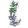
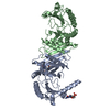
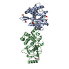
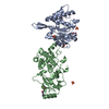
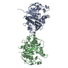
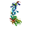
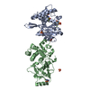
- Structure visualization
| Structure viewer | Molecule: MolmilJmol/JSmol |
|---|
- Downloads & links
-Download
| PDBx/mmCIF format | 1bo1.cif.gz | 140.6 KB | Display | PDBx/mmCIF format |
|---|---|---|---|---|
| PDB format | pdb1bo1.ent.gz | 111.5 KB | Display | PDB format |
| PDBx/mmJSON format | 1bo1.json.gz | Tree view | PDBx/mmJSON format | |
| Others |  Other downloads Other downloads |
-Validation report
| Arichive directory | https://data.pdbj.org/pub/pdb/validation_reports/bo/1bo1ftp://data.pdbj.org/pub/pdb/validation_reports/bo/1bo1 | HTTPS FTP |
|---|
-Related structure data
| Similar structure data |
|---|


-Links
 PDBj
PDBj


- Assembly
| Deposited unit | 
| ||||||||
|---|---|---|---|---|---|---|---|---|---|
| 1 |
| ||||||||
| Unit cell |
| ||||||||
| Noncrystallographic symmetry (NCS) | NCS oper: (Code: given Matrix: (-0.996792, 0.067419, -0.043137), Vector: |
-Components
| #1: Protein | Mass: 47447.926 Da / Num. of mol.: 2 Source method: isolated from a genetically manipulated source Source: (gene. exp.) Homo sapiens (human) / Description: HUMAN ENZYME CLONED INTO E.COLI. / Plasmid: PET28B / Cell line (production host): BL21(DE3) / Production host:  References: UniProt: P78356, 1-phosphatidylinositol-4-phosphate 5-kinase #2: Water | ChemComp-HOH / |  Mass: 18.015 Da / Num. of mol.: 19 / Source method: isolated from a natural source / Formula: H2O Mass: 18.015 Da / Num. of mol.: 19 / Source method: isolated from a natural source / Formula: H2O |
|---|
-Experimental details
-Experiment
| Experiment | Method: X-RAY DIFFRACTION / Number of used crystals: 2 |
|---|
- Sample preparation
| Crystal | Density Matthews: 2.6 Å3/Da / Density % sol: 54 % | ||||||||||||||||||||||||||||||||||||
|---|---|---|---|---|---|---|---|---|---|---|---|---|---|---|---|---|---|---|---|---|---|---|---|---|---|---|---|---|---|---|---|---|---|---|---|---|---|
| Crystal grow | pH: 5.6 Details: 100MM SODIUM CITRATE PH5.6 200MM MAGNESIUM ACETATE 100MM LITHIUM ACETATE 16% PEG (4000) | ||||||||||||||||||||||||||||||||||||
| Crystal grow | *PLUS Temperature: 20 ℃ / Method: vapor diffusion, hanging drop | ||||||||||||||||||||||||||||||||||||
| Components of the solutions | *PLUS
|
-Data collection
| Diffraction | Mean temperature: 300 K |
|---|---|
| Diffraction source | Source: SYNCHROTRON / Site: NSLS  / Beamline: X9B / Wavelength: 1.5418 / Beamline: X9B / Wavelength: 1.5418 |
| Detector | Type: MACSCIENCE / Detector: IMAGE PLATE / Date: Feb 15, 1998 / Details: MIRRORS |
| Radiation | Monochromator: NI FILTER / Protocol: SINGLE WAVELENGTH / Monochromatic (M) / Laue (L): M / Scattering type: x-ray |
| Radiation wavelength | Wavelength: 1.5418 Å / Relative weight: 1 |
| Reflection | Resolution: 3→20 Å / Num. obs: 21621 / % possible obs: 99.2 % / Observed criterion σ(I): 0 / Redundancy: 13.7 % / Rsym value: 0.052 / Net I/σ(I): 14.7 |
| Reflection shell | Resolution: 3→3.11 Å / Redundancy: 10.2 % / Mean I/σ(I) obs: 4.5 / Rsym value: 0.325 / % possible all: 98.3 |
| Reflection | *PLUS Num. measured all: 297963 / Rmerge(I) obs: 0.052 |
| Reflection shell | *PLUS % possible obs: 98.3 % |
- Processing
| Software |
| ||||||||||||||||||||||||||||||||||||||||||||||||||||||||||||
|---|---|---|---|---|---|---|---|---|---|---|---|---|---|---|---|---|---|---|---|---|---|---|---|---|---|---|---|---|---|---|---|---|---|---|---|---|---|---|---|---|---|---|---|---|---|---|---|---|---|---|---|---|---|---|---|---|---|---|---|---|---|
| Refinement | Method to determine structure: SIRAS / Resolution: 3→6 Å / Data cutoff high absF: 100000 / Data cutoff low absF: 0.1 / Cross valid method: THROUGHOUT / σ(F): 0
| ||||||||||||||||||||||||||||||||||||||||||||||||||||||||||||
| Displacement parameters | Biso mean: 48.05 Å2 | ||||||||||||||||||||||||||||||||||||||||||||||||||||||||||||
| Refinement step | Cycle: LAST / Resolution: 3→6 Å
| ||||||||||||||||||||||||||||||||||||||||||||||||||||||||||||
| Refine LS restraints |
| ||||||||||||||||||||||||||||||||||||||||||||||||||||||||||||
| Refine LS restraints NCS | NCS model details: RESTRAINTS | ||||||||||||||||||||||||||||||||||||||||||||||||||||||||||||
| LS refinement shell | Resolution: 3→3.12 Å / Total num. of bins used: 8
| ||||||||||||||||||||||||||||||||||||||||||||||||||||||||||||
| Xplor file |
| ||||||||||||||||||||||||||||||||||||||||||||||||||||||||||||
| Refinement | *PLUS % reflection Rfree: 5 % | ||||||||||||||||||||||||||||||||||||||||||||||||||||||||||||
| Solvent computation | *PLUS | ||||||||||||||||||||||||||||||||||||||||||||||||||||||||||||
| Displacement parameters | *PLUS | ||||||||||||||||||||||||||||||||||||||||||||||||||||||||||||
| Refine LS restraints | *PLUS
| ||||||||||||||||||||||||||||||||||||||||||||||||||||||||||||
| LS refinement shell | *PLUS % reflection Rfree: 5 % |
