 Movie
Movie Controller

[English] 日本語
 Yorodumi
- PDB-2y69: Bovine heart cytochrome c oxidase re-refined with molecular oxygen -
+ Open data

- Basic information
| Entry | Database: PDB / ID: 2y69 | ||||||
|---|---|---|---|---|---|---|---|
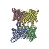
| Title | Bovine heart cytochrome c oxidase re-refined with molecular oxygen | ||||||
 Components Components |
| ||||||
 Keywords Keywords | ELECTRON TRANSPORT / COMPLEX IV / PROTON PUMPS / MEMBRANE PROTEIN | ||||||
| Function / homology |  Function and homology information Function and homology informationComplex IV assembly / TP53 Regulates Metabolic Genes / respiratory chain complex IV assembly / Cytoprotection by HMOX1 / mitochondrial respirasome assembly / Respiratory electron transport / respiratory chain complex IV / respiratory chain complex / Mitochondrial translation termination / cytochrome-c oxidase ...Complex IV assembly / TP53 Regulates Metabolic Genes / respiratory chain complex IV assembly / Cytoprotection by HMOX1 / mitochondrial respirasome assembly / Respiratory electron transport / respiratory chain complex IV / respiratory chain complex / Mitochondrial translation termination / cytochrome-c oxidase / oxidative phosphorylation / mitochondrial electron transport, cytochrome c to oxygen / cytochrome-c oxidase activity / Mitochondrial protein degradation / ATP synthesis coupled electron transport / enzyme regulator activity / proton transmembrane transport / aerobic respiration / respiratory electron transport chain / central nervous system development / mitochondrial membrane / oxidoreductase activity / mitochondrial inner membrane / copper ion binding / heme binding / mitochondrion / metal ion binding Similarity search - Function | ||||||
| Biological species |  | ||||||
| Method |  X-RAY DIFFRACTION / MOLECULAR REPLACEMENT / Resolution: 1.95 Å X-RAY DIFFRACTION / MOLECULAR REPLACEMENT / Resolution: 1.95 Å | ||||||
 Authors Authors | Kaila, V.R.I. / Oksanen, E. / Goldman, A. / Verkhovsky, M.I. / Sundholm, D. / Wikstrom, M. | ||||||
 Citation Citation | Journal: Proc.Natl.Acad.Sci.USA / Year: 2009 Title: A Peroxide Bridge between Fe and Cu Ions in the O2 Reduction Site of Fully Oxidized Cytochrome C Oxidase Could Suppress the Proton Pump. Authors: Aoyama, H. / Muramoto, K. / Shinzawa-Itoh, K. / Hirata, K. / Yamashita, E. / Tsukihara, T. / Ogura, T. / Yoshikawa, S. | ||||||
| History |
| ||||||
| Remark 0 | THIS ENTRY 2Y69 REFLECTS AN ALTERNATIVE MODELING OF THE ORIGINAL STRUCTURAL DATA (R3ABMSF) ...THIS ENTRY 2Y69 REFLECTS AN ALTERNATIVE MODELING OF THE ORIGINAL STRUCTURAL DATA (R3ABMSF) DETERMINED BY AUTHORS OF THE PDB ENTRY 3ABM: H.AOYAMA,K.MURAMOTO,K.SHINZAWA-ITOH,K.HIRATA,E.YAMASHITA, T.TSUKIHARA,T.OGURA,S.YOSHIKAWA |
- Structure visualization
| Structure viewer | Molecule: MolmilJmol/JSmol |
|---|
- Downloads & links
-Download
| PDBx/mmCIF format | 2y69.cif.gz | 763.4 KB | Display | PDBx/mmCIF format |
|---|---|---|---|---|
| PDB format | pdb2y69.ent.gz | 618.2 KB | Display | PDB format |
| PDBx/mmJSON format | 2y69.json.gz | Tree view | PDBx/mmJSON format | |
| Others |  Other downloads Other downloads |
-Validation report
| Arichive directory | https://data.pdbj.org/pub/pdb/validation_reports/y6/2y69ftp://data.pdbj.org/pub/pdb/validation_reports/y6/2y69 | HTTPS FTP |
|---|
-Related structure data
| Related structure data | |
|---|---|
| Similar structure data |


-Links
 PDBj
PDBj


- Assembly
| Deposited unit | 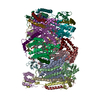
| ||||||||
|---|---|---|---|---|---|---|---|---|---|
| 1 | 
| ||||||||
| 2 | 
| ||||||||
| Unit cell |
|
-Components
-CYTOCHROME C OXIDASE SUBUNIT ... , 8 types, 16 molecules ANBOCPDQERFSHULY
| #1: Protein | Mass: 57065.844 Da / Num. of mol.: 2 / Source method: isolated from a natural source / Source: (natural) #2: Protein | Mass: 26040.393 Da / Num. of mol.: 2 / Source method: isolated from a natural source / Source: (natural) #3: Protein | Mass: 29957.627 Da / Num. of mol.: 2 / Source method: isolated from a natural source / Source: (natural) #4: Protein | Mass: 19602.609 Da / Num. of mol.: 2 / Source method: isolated from a natural source / Source: (natural) #5: Protein | Mass: 16758.156 Da / Num. of mol.: 2 / Source method: isolated from a natural source / Source: (natural) #6: Protein | Mass: 13852.807 Da / Num. of mol.: 2 / Source method: isolated from a natural source / Source: (natural) #8: Protein | Mass: 10170.439 Da / Num. of mol.: 2 / Source method: isolated from a natural source / Source: (natural) #12: Protein | Mass: 7342.661 Da / Num. of mol.: 2 / Source method: isolated from a natural source / Source: (natural) |
|---|
-CYTOCHROME C OXIDASE POLYPEPTIDE ... , 5 types, 10 molecules GTIVJWKXMZ
| #7: Protein | Mass: 10899.382 Da / Num. of mol.: 2 / Source method: isolated from a natural source / Source: (natural) #9: Protein | Mass: 8626.179 Da / Num. of mol.: 2 / Source method: isolated from a natural source / Source: (natural) #10: Protein | Mass: 9076.522 Da / Num. of mol.: 2 / Source method: isolated from a natural source / Source: (natural) #11: Protein | Mass: 9077.439 Da / Num. of mol.: 2 / Source method: isolated from a natural source / Source: (natural) #13: Protein | Mass: 7650.059 Da / Num. of mol.: 2 / Source method: isolated from a natural source / Source: (natural) |
|---|
-Sugars , 1 types, 2 molecules 
| #23: Sugar |  Type: D-saccharide / Mass: 482.562 Da / Num. of mol.: 2 Type: D-saccharide / Mass: 482.562 Da / Num. of mol.: 2Source method: isolated from a genetically manipulated source Formula: C22H42O11 / Comment: detergent*YM |
|---|
-Non-polymers , 10 types, 1074 molecules 


| #14: Chemical | ChemComp-HEA /  Mass: 852.837 Da / Num. of mol.: 4 / Source method: obtained synthetically / Formula: C49H56FeN4O6 Mass: 852.837 Da / Num. of mol.: 4 / Source method: obtained synthetically / Formula: C49H56FeN4O6#15: Chemical |  Mass: 63.546 Da / Num. of mol.: 2 / Source method: obtained synthetically / Formula: Cu Mass: 63.546 Da / Num. of mol.: 2 / Source method: obtained synthetically / Formula: Cu#16: Chemical |  Mass: 31.999 Da / Num. of mol.: 2 / Source method: obtained synthetically / Formula: O2 Mass: 31.999 Da / Num. of mol.: 2 / Source method: obtained synthetically / Formula: O2#17: Chemical |  Mass: 24.305 Da / Num. of mol.: 2 / Source method: obtained synthetically / Formula: Mg Mass: 24.305 Da / Num. of mol.: 2 / Source method: obtained synthetically / Formula: Mg#18: Chemical | ChemComp-CHD /  Mass: 408.571 Da / Num. of mol.: 6 / Source method: obtained synthetically / Formula: C24H40O5 Mass: 408.571 Da / Num. of mol.: 6 / Source method: obtained synthetically / Formula: C24H40O5#19: Chemical |  Mass: 127.092 Da / Num. of mol.: 2 / Source method: obtained synthetically / Formula: Cu2 Mass: 127.092 Da / Num. of mol.: 2 / Source method: obtained synthetically / Formula: Cu2#20: Chemical |  Mass: 768.055 Da / Num. of mol.: 2 / Source method: obtained synthetically / Formula: C43H78NO8P / Comment: phospholipid*YM Mass: 768.055 Da / Num. of mol.: 2 / Source method: obtained synthetically / Formula: C43H78NO8P / Comment: phospholipid*YM#21: Chemical | ChemComp-PGV / (  Mass: 749.007 Da / Num. of mol.: 4 / Source method: obtained synthetically / Formula: C40H77O10P / Comment: phospholipid*YM Mass: 749.007 Da / Num. of mol.: 4 / Source method: obtained synthetically / Formula: C40H77O10P / Comment: phospholipid*YM#22: Chemical |  Mass: 65.409 Da / Num. of mol.: 2 / Source method: obtained synthetically / Formula: Zn Mass: 65.409 Da / Num. of mol.: 2 / Source method: obtained synthetically / Formula: Zn#24: Water | ChemComp-HOH / | Mass: 18.015 Da / Num. of mol.: 1048 / Source method: isolated from a natural source / Formula: H2O |
|---|
-Details
| Has protein modification | Y |
|---|
-Experimental details
-Experiment
| Experiment | Method: X-RAY DIFFRACTION / Number of used crystals: 1 |
|---|
- Sample preparation
| Crystal | Density Matthews: 3.75 Å3/Da / Density % sol: 67.17 % / Description: AUTHOR USED THE SF DATA FROM ENTRY 3ABM |
|---|
-Data collection
| Radiation | Protocol: SINGLE WAVELENGTH / Monochromatic (M) / Laue (L): M / Scattering type: x-ray |
|---|---|
| Radiation wavelength | Relative weight: 1 |
| Reflection | Biso Wilson estimate: 0 Å2 |
- Processing
| Software | Name: REFMAC / Version: 5.5.0066 / Classification: refinement | ||||||||||||||||||||||||||||||||||||||||||||||||||||||||||||||||||||||||||||||||||||||||||||||||||||||||||||||||||||||||||||||||||||||||||||||||||||||||||||||||||||||||||||||||||||||
|---|---|---|---|---|---|---|---|---|---|---|---|---|---|---|---|---|---|---|---|---|---|---|---|---|---|---|---|---|---|---|---|---|---|---|---|---|---|---|---|---|---|---|---|---|---|---|---|---|---|---|---|---|---|---|---|---|---|---|---|---|---|---|---|---|---|---|---|---|---|---|---|---|---|---|---|---|---|---|---|---|---|---|---|---|---|---|---|---|---|---|---|---|---|---|---|---|---|---|---|---|---|---|---|---|---|---|---|---|---|---|---|---|---|---|---|---|---|---|---|---|---|---|---|---|---|---|---|---|---|---|---|---|---|---|---|---|---|---|---|---|---|---|---|---|---|---|---|---|---|---|---|---|---|---|---|---|---|---|---|---|---|---|---|---|---|---|---|---|---|---|---|---|---|---|---|---|---|---|---|---|---|---|---|
| Refinement | Method to determine structure: MOLECULAR REPLACEMENT / Resolution: 1.95→64.15 Å / Cor.coef. Fo:Fc: 0.95 / Cor.coef. Fo:Fc free: 0.926 / SU B: 3.281 / SU ML: 0.093 / Cross valid method: THROUGHOUT / ESU R: 0.125 / ESU R Free: 0.125 / Stereochemistry target values: MAXIMUM LIKELIHOOD Details: HYDROGENS HAVE BEEN ADDED IN THE RIDING POSITIONS. THIS ENTRY REFLECTS AN ALTERNATIVE MODELING OF X-RAY DATA R3ABMSF.
| ||||||||||||||||||||||||||||||||||||||||||||||||||||||||||||||||||||||||||||||||||||||||||||||||||||||||||||||||||||||||||||||||||||||||||||||||||||||||||||||||||||||||||||||||||||||
| Solvent computation | Ion probe radii: 0.8 Å / Shrinkage radii: 0.8 Å / VDW probe radii: 1.4 Å / Solvent model: MASK | ||||||||||||||||||||||||||||||||||||||||||||||||||||||||||||||||||||||||||||||||||||||||||||||||||||||||||||||||||||||||||||||||||||||||||||||||||||||||||||||||||||||||||||||||||||||
| Displacement parameters | Biso mean: 45.538 Å2
| ||||||||||||||||||||||||||||||||||||||||||||||||||||||||||||||||||||||||||||||||||||||||||||||||||||||||||||||||||||||||||||||||||||||||||||||||||||||||||||||||||||||||||||||||||||||
| Refinement step | Cycle: LAST / Resolution: 1.95→64.15 Å
| ||||||||||||||||||||||||||||||||||||||||||||||||||||||||||||||||||||||||||||||||||||||||||||||||||||||||||||||||||||||||||||||||||||||||||||||||||||||||||||||||||||||||||||||||||||||
| Refine LS restraints |
|