 Movie
Movie Controller


+ Open data

- Basic information
| Entry | Database: PDB / ID: 2y4t | ||||||
|---|---|---|---|---|---|---|---|
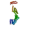
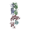
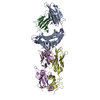
| Title | Crystal structure of the human co-chaperone P58(IPK) | ||||||
 Components Components | DNAJ HOMOLOG SUBFAMILY C MEMBER 3 | ||||||
 Keywords Keywords | CHAPERONE / ENDOPLASMIC RETICULUM / PROTEIN FOLDING / TETRATRICOPEPTIDEREPEAT / J DOMAIN / UNFOLDED PROTEIN RESPONSE | ||||||
| Function / homology |  Function and homology information Function and homology informationpositive regulation of translation initiation in response to endoplasmic reticulum stress / negative regulation of endoplasmic reticulum stress-induced eIF2 alpha phosphorylation / negative regulation of translation in response to endoplasmic reticulum stress / protein folding in endoplasmic reticulum / XBP1(S) activates chaperone genes / misfolded protein binding / cellular response to cold / protein kinase inhibitor activity / response to unfolded protein / Viral mRNA Translation ...positive regulation of translation initiation in response to endoplasmic reticulum stress / negative regulation of endoplasmic reticulum stress-induced eIF2 alpha phosphorylation / negative regulation of translation in response to endoplasmic reticulum stress / protein folding in endoplasmic reticulum / XBP1(S) activates chaperone genes / misfolded protein binding / cellular response to cold / protein kinase inhibitor activity / response to unfolded protein / Viral mRNA Translation / : / Post-translational protein phosphorylation / PKR-mediated signaling / Regulation of Insulin-like Growth Factor (IGF) transport and uptake by Insulin-like Growth Factor Binding Proteins (IGFBPs) / azurophil granule lumen / extracellular vesicle / protein-folding chaperone binding / defense response to virus / endoplasmic reticulum lumen / Neutrophil degranulation / protein kinase binding / negative regulation of apoptotic process / endoplasmic reticulum / extracellular exosome / extracellular region / membrane / cytoplasm / cytosol Similarity search - Function | ||||||
| Biological species |  HOMO SAPIENS (human) HOMO SAPIENS (human) | ||||||
| Method |  X-RAY DIFFRACTION / SYNCHROTRON / MOLECULAR REPLACEMENT / Resolution: 3 Å X-RAY DIFFRACTION / SYNCHROTRON / MOLECULAR REPLACEMENT / Resolution: 3 Å | ||||||
 Authors Authors | Svard, M. / Biterova, E.I. / Bourhis, J.-M. / Guy, J.E. | ||||||
 Citation Citation | Journal: Plos One / Year: 2011 Title: The Crystal Structure of the Human Co-Chaperone P58Ipk Authors: Svard, M. / Biterova, E.I. / Bourhis, J.-M. / Guy, J.E. | ||||||
| History |
|
- Structure visualization
| Structure viewer | Molecule: MolmilJmol/JSmol |
|---|
- Downloads & links
-Download
| PDBx/mmCIF format | 2y4t.cif.gz | 506.8 KB | Display | PDBx/mmCIF format |
|---|---|---|---|---|
| PDB format | pdb2y4t.ent.gz | 426.1 KB | Display | PDB format |
| PDBx/mmJSON format | 2y4t.json.gz | Tree view | PDBx/mmJSON format | |
| Others |  Other downloads Other downloads |
-Validation report
| Arichive directory | https://data.pdbj.org/pub/pdb/validation_reports/y4/2y4tftp://data.pdbj.org/pub/pdb/validation_reports/y4/2y4t | HTTPS FTP |
|---|
-Related structure data

-Links
 PDBj
PDBj


- Assembly
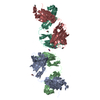
| Deposited unit | 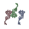
| ||||||||||||||||
|---|---|---|---|---|---|---|---|---|---|---|---|---|---|---|---|---|---|
| 1 | 
| ||||||||||||||||
| 2 | 
| ||||||||||||||||
| 3 | 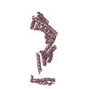
| ||||||||||||||||
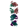
| Unit cell |
| ||||||||||||||||
| Components on special symmetry positions |
| ||||||||||||||||
| Noncrystallographic symmetry (NCS) | NCS oper:
|
-Components
| #1: Protein | Mass: 51946.836 Da / Num. of mol.: 3 / Fragment: RESIDUES 35-461 Source method: isolated from a genetically manipulated source Source: (gene. exp.) HOMO SAPIENS (human) / Plasmid: P58IPK_6 / Production host:  #2: Water | ChemComp-HOH / |  Mass: 18.015 Da / Num. of mol.: 25 / Source method: isolated from a natural source / Formula: H2O Mass: 18.015 Da / Num. of mol.: 25 / Source method: isolated from a natural source / Formula: H2OHas protein modification | Y | |
|---|
-Experimental details
-Experiment
| Experiment | Method: X-RAY DIFFRACTION |
|---|
- Sample preparation
| Crystal | Density Matthews: 3.9 Å3/Da / Density % sol: 69 % / Description: NONE |
|---|---|
| Crystal grow | Details: 0.1 M HEPES, PH 8.2, 1.4 M SUCCINIC ACID, 1% (W/V) PEG MME 2000 |
-Data collection
| Diffraction | Mean temperature: 100 K |
|---|---|
| Diffraction source | Source: SYNCHROTRON / Site: ESRF  / Beamline: ID14-1 / Wavelength: 0.933 / Beamline: ID14-1 / Wavelength: 0.933 |
| Detector | Type: ADSC CCD / Detector: CCD |
| Radiation | Protocol: SINGLE WAVELENGTH / Monochromatic (M) / Laue (L): M / Scattering type: x-ray |
| Radiation wavelength | Wavelength: 0.933 Å / Relative weight: 1 |
| Reflection | Resolution: 3→25 Å / Num. obs: 44987 / % possible obs: 96.6 % / Observed criterion σ(I): 2 / Redundancy: 4.3 % / Biso Wilson estimate: 74.9 Å2 / Rmerge(I) obs: 0.08 / Net I/σ(I): 11.5 |
| Reflection shell | Resolution: 3→3.08 Å / Redundancy: 3.7 % / Rmerge(I) obs: 0.56 / Mean I/σ(I) obs: 2.2 / % possible all: 79.2 |
- Processing
| Software |
| ||||||||||||||||||||||||||||||||||||||||||||||||||||||||||||||||||||||||||||||||||||||||||||||||||||||||||||||||||||||||||||||||||||||||||||||||||||||||||||||||||||||||||||||||||||||
|---|---|---|---|---|---|---|---|---|---|---|---|---|---|---|---|---|---|---|---|---|---|---|---|---|---|---|---|---|---|---|---|---|---|---|---|---|---|---|---|---|---|---|---|---|---|---|---|---|---|---|---|---|---|---|---|---|---|---|---|---|---|---|---|---|---|---|---|---|---|---|---|---|---|---|---|---|---|---|---|---|---|---|---|---|---|---|---|---|---|---|---|---|---|---|---|---|---|---|---|---|---|---|---|---|---|---|---|---|---|---|---|---|---|---|---|---|---|---|---|---|---|---|---|---|---|---|---|---|---|---|---|---|---|---|---|---|---|---|---|---|---|---|---|---|---|---|---|---|---|---|---|---|---|---|---|---|---|---|---|---|---|---|---|---|---|---|---|---|---|---|---|---|---|---|---|---|---|---|---|---|---|---|---|
| Refinement | Method to determine structure: MOLECULAR REPLACEMENT / Resolution: 3→25 Å / Cor.coef. Fo:Fc: 0.935 / Cor.coef. Fo:Fc free: 0.912 / SU B: 39.464 / SU ML: 0.325 / Cross valid method: THROUGHOUT / ESU R: 1.211 / ESU R Free: 0.429 / Stereochemistry target values: MAXIMUM LIKELIHOOD Details: HYDROGENS HAVE BEEN ADDED IN THE RIDING POSITIONS. HYDROGENS HAVE BEEN USED IF PRESENT IN THE INPUT. THE REGIONS THAT CORRESPOND TO AMINO ACIDS 400-406, 451-452 OF CHAIN B AND 400-409, 449- ...Details: HYDROGENS HAVE BEEN ADDED IN THE RIDING POSITIONS. HYDROGENS HAVE BEEN USED IF PRESENT IN THE INPUT. THE REGIONS THAT CORRESPOND TO AMINO ACIDS 400-406, 451-452 OF CHAIN B AND 400-409, 449-450 OF CHAIN C ARE LOCALIZED TO VERY FLEXIBLE LOOPS, CONTAIN VERY WEAK ELECTRON DENSITY AND HAVE BEEN MODELED BASED ON THE POSITION OF RESPECTIVE AMINO ACIDS OF BETTER ORDERED CHAIN A. IN CHAINS B AND C, THE LOOP BETWEEN HELICES I AND II OF THE J DOMAIN (AMINO ACID RESIDUES 402-408) AND THE LOOP IN THE J DOMAIN (RESIDUES 425-428) HAVE VERY WEAK DENSITY AND ARE MODELED BASED ON THEIR POSITION IN CHAIN A.
| ||||||||||||||||||||||||||||||||||||||||||||||||||||||||||||||||||||||||||||||||||||||||||||||||||||||||||||||||||||||||||||||||||||||||||||||||||||||||||||||||||||||||||||||||||||||
| Solvent computation | Ion probe radii: 0.8 Å / Shrinkage radii: 0.8 Å / VDW probe radii: 1.2 Å / Solvent model: MASK | ||||||||||||||||||||||||||||||||||||||||||||||||||||||||||||||||||||||||||||||||||||||||||||||||||||||||||||||||||||||||||||||||||||||||||||||||||||||||||||||||||||||||||||||||||||||
| Displacement parameters | Biso mean: 107.121 Å2
| ||||||||||||||||||||||||||||||||||||||||||||||||||||||||||||||||||||||||||||||||||||||||||||||||||||||||||||||||||||||||||||||||||||||||||||||||||||||||||||||||||||||||||||||||||||||
| Refinement step | Cycle: LAST / Resolution: 3→25 Å
| ||||||||||||||||||||||||||||||||||||||||||||||||||||||||||||||||||||||||||||||||||||||||||||||||||||||||||||||||||||||||||||||||||||||||||||||||||||||||||||||||||||||||||||||||||||||
| Refine LS restraints |
|