 Movie
Movie Controller

[English] 日本語
 Yorodumi
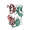
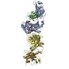
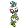
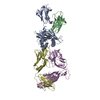
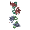
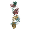
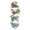
- PDB-1pg7: Murine 6A6 Fab in complex with humanized anti-Tissue Factor D3H44 Fab -
+ Open data

- Basic information
| Entry | Database: PDB / ID: 1pg7 | ||||||
|---|---|---|---|---|---|---|---|
| Title | Murine 6A6 Fab in complex with humanized anti-Tissue Factor D3H44 Fab | ||||||
 Components Components |
| ||||||
 Keywords Keywords | IMMUNE SYSTEM | ||||||
| Function / homology |  Function and homology information Function and homology informationcomplement-dependent cytotoxicity / antibody-dependent cellular cytotoxicity / Fc-gamma receptor I complex binding / immunoglobulin complex, circulating / Classical antibody-mediated complement activation / immunoglobulin receptor binding / Initial triggering of complement / IgG immunoglobulin complex / FCGR activation / complement activation, classical pathway ...complement-dependent cytotoxicity / antibody-dependent cellular cytotoxicity / Fc-gamma receptor I complex binding / immunoglobulin complex, circulating / Classical antibody-mediated complement activation / immunoglobulin receptor binding / Initial triggering of complement / IgG immunoglobulin complex / FCGR activation / complement activation, classical pathway / Role of phospholipids in phagocytosis / immunoglobulin complex / antigen binding / FCGR3A-mediated IL10 synthesis / Regulation of Complement cascade / B cell receptor signaling pathway / FCGR3A-mediated phagocytosis / Regulation of actin dynamics for phagocytic cup formation / antibacterial humoral response / Interleukin-4 and Interleukin-13 signaling / blood microparticle / adaptive immune response / : / extracellular exosome / extracellular region / plasma membrane Similarity search - Function | ||||||
| Biological species |   Homo sapiens (human) Homo sapiens (human) | ||||||
| Method |  X-RAY DIFFRACTION / SYNCHROTRON / MOLECULAR REPLACEMENT / Resolution: 2.5 Å X-RAY DIFFRACTION / SYNCHROTRON / MOLECULAR REPLACEMENT / Resolution: 2.5 Å | ||||||
 Authors Authors | Eigenbrot, C. / Meng, Y.G. / Krishnamurthy, R. / Lipari, M.T. / Presta, L. / Devaux, B. / Wong, T. / Moran, P. / Bullens, S. / Kirchhofer, D. | ||||||
 Citation Citation | Journal: J.Mol.Biol. / Year: 2003 Title: Structural insight into how an anti-idiotypic antibody against D3H44 (anti-tissue factor antibody) restores normal coagulation. Authors: Eigenbrot, C. / Meng, Y.G. / Krishnamurthy, R. / Lipari, M.T. / Presta, L. / Devaux, B. / Wong, T. / Moran, P. / Bullens, S. / Kirchhofer, D. | ||||||
| History |
| ||||||
| Remark 600 | HETEROGEN SOLVENT ATOMS NUMBERED 1 - 125 ARE ASSOCIATED WITH CHAINS L, H, W, AND/OR X. SOLVENT ...HETEROGEN SOLVENT ATOMS NUMBERED 1 - 125 ARE ASSOCIATED WITH CHAINS L, H, W, AND/OR X. SOLVENT ATOMS NUMBERED 601 - 749 ARE WITH CHAINS M, I, Y AND/OR Z. |
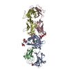
- Structure visualization
| Structure viewer | Molecule: MolmilJmol/JSmol |
|---|
- Downloads & links
-Download
| PDBx/mmCIF format | 1pg7.cif.gz | 336.7 KB | Display | PDBx/mmCIF format |
|---|---|---|---|---|
| PDB format | pdb1pg7.ent.gz | 270.6 KB | Display | PDB format |
| PDBx/mmJSON format | 1pg7.json.gz | Tree view | PDBx/mmJSON format | |
| Others |  Other downloads Other downloads |
-Validation report
| Arichive directory | https://data.pdbj.org/pub/pdb/validation_reports/pg/1pg7ftp://data.pdbj.org/pub/pdb/validation_reports/pg/1pg7 | HTTPS FTP |
|---|
-Related structure data


-Links
 PDBj
PDBj


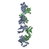
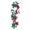
- Assembly
| Deposited unit | 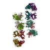
| ||||||||
|---|---|---|---|---|---|---|---|---|---|
| 1 | 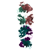
| ||||||||
| 2 | 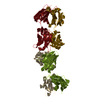
| ||||||||
| Unit cell |
|
-Components
| #1: Antibody | Mass: 23139.779 Da / Num. of mol.: 2 / Fragment: antigen-binding fragment Source method: isolated from a genetically manipulated source Source: (gene. exp.) Mus musculus, Homo sapiens / Production host:  #2: Antibody | Mass: 23419.965 Da / Num. of mol.: 2 / Fragment: antigen-binding fragment Source method: isolated from a genetically manipulated source Source: (gene. exp.) Mus musculus, Homo sapiens / Production host: #3: Antibody | Mass: 22481.793 Da / Num. of mol.: 2 / Source method: isolated from a natural source / Source: (natural) #4: Antibody | Mass: 23815.732 Da / Num. of mol.: 2 / Source method: isolated from a natural source / Source: (natural) #5: Water | ChemComp-HOH / |  Mass: 18.015 Da / Num. of mol.: 274 / Source method: isolated from a natural source / Formula: H2O Mass: 18.015 Da / Num. of mol.: 274 / Source method: isolated from a natural source / Formula: H2OHas protein modification | Y | |
|---|
-Experimental details
-Experiment
| Experiment | Method: X-RAY DIFFRACTION / Number of used crystals: 1 |
|---|
- Sample preparation
| Crystal | Density Matthews: 2.89 Å3/Da / Density % sol: 57.49 % | ||||||||||||||||||||||||||||||
|---|---|---|---|---|---|---|---|---|---|---|---|---|---|---|---|---|---|---|---|---|---|---|---|---|---|---|---|---|---|---|---|
| Crystal grow | Temperature: 277 K / Method: vapor diffusion, hanging drop / pH: 7 Details: PEG 3350, pH 7.0, VAPOR DIFFUSION, HANGING DROP, temperature 277K | ||||||||||||||||||||||||||||||
| Crystal grow | *PLUS Temperature: 4 ℃ / pH: 8 / Method: vapor diffusion, hanging drop | ||||||||||||||||||||||||||||||
| Components of the solutions | *PLUS
|
-Data collection
| Diffraction | Mean temperature: 100 K |
|---|---|
| Diffraction source | Source: SYNCHROTRON / Site: APS  / Beamline: 19-ID / Wavelength: 0.917 Å / Beamline: 19-ID / Wavelength: 0.917 Å |
| Detector | Type: CUSTOM-MADE / Detector: CCD / Date: Dec 21, 2001 |
| Radiation | Protocol: SINGLE WAVELENGTH / Monochromatic (M) / Laue (L): M / Scattering type: x-ray |
| Radiation wavelength | Wavelength: 0.917 Å / Relative weight: 1 |
| Reflection | Resolution: 2.5→50 Å / Num. all: 72248 / Num. obs: 72248 / % possible obs: 99 % / Observed criterion σ(F): 0 / Observed criterion σ(I): 0 / Redundancy: 4 % / Biso Wilson estimate: 52.4 Å2 / Rmerge(I) obs: 0.129 / Net I/σ(I): 12 |
| Reflection shell | Resolution: 2.5→2.59 Å / Redundancy: 4 % / Rmerge(I) obs: 0.477 / Num. unique all: 7157 / % possible all: 98 |
| Reflection | *PLUS Num. measured all: 310288 |
| Reflection shell | *PLUS % possible obs: 98 % / Num. unique obs: 7157 / Num. measured obs: 26775 / Mean I/σ(I) obs: 2.5 |
- Processing
| Software |
| ||||||||||||||||||||||||||||||||||||
|---|---|---|---|---|---|---|---|---|---|---|---|---|---|---|---|---|---|---|---|---|---|---|---|---|---|---|---|---|---|---|---|---|---|---|---|---|---|
| Refinement | Method to determine structure: MOLECULAR REPLACEMENT Starting model: PDB entries 1JPS, 1GIG Resolution: 2.5→50 Å / Rfactor Rfree error: 0.006 / Data cutoff high absF: 1000000 / Data cutoff low absF: 0.001 / Isotropic thermal model: RESTRAINED / Cross valid method: THROUGHOUT / σ(F): 0.2 / Stereochemistry target values: Engh & Huber / Details: BULK SOLVENT MODEL USED
| ||||||||||||||||||||||||||||||||||||
| Displacement parameters | Biso mean: 34.1 Å2
| ||||||||||||||||||||||||||||||||||||
| Refine analyze |
| ||||||||||||||||||||||||||||||||||||
| Refinement step | Cycle: LAST / Resolution: 2.5→50 Å
| ||||||||||||||||||||||||||||||||||||
| Refine LS restraints |
| ||||||||||||||||||||||||||||||||||||
| LS refinement shell | Resolution: 2.5→2.59 Å / Rfactor Rfree error: 0.028 / Total num. of bins used: 10
| ||||||||||||||||||||||||||||||||||||
| Xplor file |
| ||||||||||||||||||||||||||||||||||||
| Software | *PLUS Name: X-PLOR / Classification: refinement | ||||||||||||||||||||||||||||||||||||
| Refinement | *PLUS Num. reflection Rfree: 2193 / % reflection Rfree: 3 % / Rfactor Rfree: 0.267 / Rfactor Rwork: 0.219 | ||||||||||||||||||||||||||||||||||||
| Solvent computation | *PLUS | ||||||||||||||||||||||||||||||||||||
| Displacement parameters | *PLUS | ||||||||||||||||||||||||||||||||||||
| Refine LS restraints | *PLUS
| ||||||||||||||||||||||||||||||||||||
| LS refinement shell | *PLUS Rfactor Rfree: 0.411 / Rfactor Rwork: 0.341 |
