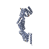
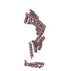
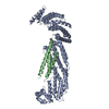
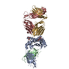
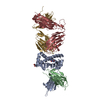
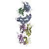
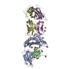
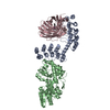
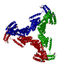
Entry Database : PDB / ID : 2y4uTitle Crystal structure of human P58(IPK) in space group P312 DNAJ HOMOLOG SUBFAMILY C MEMBER 3 Keywords / / / / / Function / homology Function Domain/homology Component
/ / / / / / / / / / / / / / / / / / / / / / / / / / / / / / / / / / / / / / / / / / / / / / / / / / / / / / / / / / / / / / / / / / / Biological species HOMO SAPIENS (human)Method / / / Resolution : 3.2 Å Authors Svard, M. / Biterova, E.I. / Bourhis, J.-M. / Guy, J.E. Journal : Plos One / Year : 2011Title : The Crystal Structure of the Human Co-Chaperone P58IpkAuthors : Svard, M. / Biterova, E.I. / Bourhis, J. / Guy, J.E. History Deposition Jan 10, 2011 Deposition site / Processing site Revision 1.0 Aug 10, 2011 Provider / Type Revision 1.1 Dec 20, 2023 Group Data collection / Database references ... Data collection / Database references / Derived calculations / Other / Refinement description Category chem_comp_atom / chem_comp_bond ... chem_comp_atom / chem_comp_bond / database_2 / pdbx_database_status / pdbx_initial_refinement_model / struct_conn Item _database_2.pdbx_DOI / _database_2.pdbx_database_accession ... _database_2.pdbx_DOI / _database_2.pdbx_database_accession / _pdbx_database_status.status_code_sf / _struct_conn.pdbx_leaving_atom_flag Revision 1.2 Nov 6, 2024 Group / Category / pdbx_modification_feature
Show all Show less
 Movie
Movie Controller


 Open data
 Basic information
 Components
 Keywords
 Function and homology information
 HOMO SAPIENS (human)
 X-RAY DIFFRACTION /
  Authors
 Citation
 Structure visualization
 Downloads & links
 Other downloads
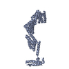


 PDBj
PDBj


 Assembly
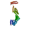

 Sample preparation
 / Beamline: ID23-1 / Wavelength: 0.979
  Processing