 Movie
Movie Controller


+ Open data

- Basic information
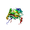
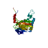
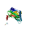
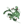
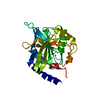
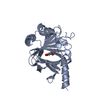
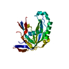
| Entry | Database: PDB / ID: 2y34 | ||||||
|---|---|---|---|---|---|---|---|
| Title | S-nitrosylated PHD2 (NO exposed) in complex with Fe(II) and UN9 | ||||||
 Components Components | EGL NINE HOMOLOG 1 | ||||||
 Keywords Keywords | OXIDOREDUCTASE / NON-HEME / TRANSCRIPTION AND EPIGENETIC REGULATION | ||||||
| Function / homology |  Function and homology information Function and homology informationnegative regulation of hypoxia-inducible factor-1alpha signaling pathway / peptidyl-proline 4-dioxygenase activity / hypoxia-inducible factor-proline dioxygenase / hypoxia-inducible factor-proline dioxygenase activity / peptidyl-proline dioxygenase activity / regulation protein catabolic process at postsynapse / intracellular oxygen homeostasis / regulation of modification of postsynaptic structure / 2-oxoglutarate-dependent dioxygenase activity / L-ascorbic acid binding ...negative regulation of hypoxia-inducible factor-1alpha signaling pathway / peptidyl-proline 4-dioxygenase activity / hypoxia-inducible factor-proline dioxygenase / hypoxia-inducible factor-proline dioxygenase activity / peptidyl-proline dioxygenase activity / regulation protein catabolic process at postsynapse / intracellular oxygen homeostasis / regulation of modification of postsynaptic structure / 2-oxoglutarate-dependent dioxygenase activity / L-ascorbic acid binding / response to nitric oxide / regulation of angiogenesis / regulation of neuron apoptotic process / enzyme inhibitor activity / ferrous iron binding / Oxygen-dependent proline hydroxylation of Hypoxia-inducible Factor Alpha / cellular response to hypoxia / intracellular iron ion homeostasis / response to hypoxia / postsynaptic density / glutamatergic synapse / enzyme binding / positive regulation of transcription by RNA polymerase II / zinc ion binding / nucleus / cytosol / cytoplasm Similarity search - Function | ||||||
| Biological species |  Homo sapiens (human) Homo sapiens (human) | ||||||
| Method |  X-RAY DIFFRACTION / SYNCHROTRON / MOLECULAR REPLACEMENT / Resolution: 2.01 Å X-RAY DIFFRACTION / SYNCHROTRON / MOLECULAR REPLACEMENT / Resolution: 2.01 Å | ||||||
 Authors Authors | Chowdhury, R. / McDonough, M.A. / Schofield, C.J. | ||||||
 Citation Citation | Journal: J. Mol. Biol. / Year: 2011 Title: Studies on the reaction of nitric oxide with the hypoxia-inducible factor prolyl hydroxylase domain 2 (EGLN1). Authors: Chowdhury, R. / Flashman, E. / Mecinovic, J. / Kramer, H.B. / Kessler, B.M. / Frapart, Y.M. / Boucher, J.L. / Clifton, I.J. / McDonough, M.A. / Schofield, C.J. #1: Journal: Structure / Year: 2009Title: Structural Basis for Binding of Hypoxia-Inducible Factor to the Oxygen-Sensing Prolyl Hydroxylases. Authors: Chowdhury, R. / McDonough, M.A. / Mecinovic, J. / Loenarz, C. / Flashman, E. / Hewitson, K.S. / Domene, C. / Schofield, C.J. #2: Journal: Proc.Natl.Acad.Sci.USA / Year: 2006Title: Cellular Oxygen Sensing: Crystal Structure of Hypoxia-Inducible Factor Prolyl Hydroxylase (Phd2). Authors: McDonough, M.A. / Li, V. / Flashman, E. / Chowdhury, R. / Mohr, C. / Lienard, B.M.R. / Zondlo, J. / Oldham, N.J. / Clifton, I.J. / Lewis, J. / Mcneill, L.A. / Kurzeja, R.J.M. / Hewitson, K.S. ...Authors: McDonough, M.A. / Li, V. / Flashman, E. / Chowdhury, R. / Mohr, C. / Lienard, B.M.R. / Zondlo, J. / Oldham, N.J. / Clifton, I.J. / Lewis, J. / Mcneill, L.A. / Kurzeja, R.J.M. / Hewitson, K.S. / Yang, E. / Jordan, S. / Syed, R.S. / Schofield, C.J. | ||||||
| History |
|
- Structure visualization
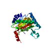
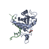
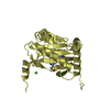
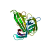
| Structure viewer | Molecule: MolmilJmol/JSmol |
|---|
- Downloads & links
-Download
| PDBx/mmCIF format | 2y34.cif.gz | 63 KB | Display | PDBx/mmCIF format |
|---|---|---|---|---|
| PDB format | pdb2y34.ent.gz | 44.6 KB | Display | PDB format |
| PDBx/mmJSON format | 2y34.json.gz | Tree view | PDBx/mmJSON format | |
| Others |  Other downloads Other downloads |
-Validation report
| Arichive directory | https://data.pdbj.org/pub/pdb/validation_reports/y3/2y34ftp://data.pdbj.org/pub/pdb/validation_reports/y3/2y34 | HTTPS FTP |
|---|
-Related structure data
| Related structure data | 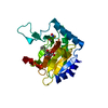 2y33C  2g19S S: Starting model for refinement C: citing same article ( |
|---|---|
| Similar structure data |


-Links
 PDBj
PDBj


- Assembly
| Deposited unit | 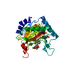
| ||||||||
|---|---|---|---|---|---|---|---|---|---|
| 1 |
| ||||||||
| Unit cell |
|
-Components
| #1: Protein | Mass: 28125.939 Da / Num. of mol.: 1 / Fragment: CATALYTIC DOMAIN, RESIDUES 181-426 Source method: isolated from a genetically manipulated source Source: (gene. exp.) Homo sapiens (human) / Production host: Escherichia coli BL21(DE3)References: UniProt: Q9GZT9, Oxidoreductases; Acting on paired donors, with incorporation or reduction of molecular oxygen; With 2-oxoglutarate as one donor, and incorporation of one atom of oxygen into each donor | ||||
|---|---|---|---|---|---|
| #2: Chemical | ChemComp-FE2 /   Mass: 55.845 Da / Num. of mol.: 1 / Source method: obtained synthetically / Formula: Fe Mass: 55.845 Da / Num. of mol.: 1 / Source method: obtained synthetically / Formula: Fe | ||||
| #3: Chemical | 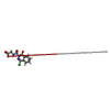  Mass: 280.664 Da / Num. of mol.: 2 / Source method: obtained synthetically / Formula: C12H9ClN2O4 Mass: 280.664 Da / Num. of mol.: 2 / Source method: obtained synthetically / Formula: C12H9ClN2O4#4: Water | ChemComp-HOH / |  Mass: 18.015 Da / Num. of mol.: 113 / Source method: isolated from a natural source / Formula: H2O Mass: 18.015 Da / Num. of mol.: 113 / Source method: isolated from a natural source / Formula: H2OHas protein modification | Y | |
-Experimental details
-Experiment
| Experiment | Method: X-RAY DIFFRACTION / Number of used crystals: 1 |
|---|
- Sample preparation
| Crystal | Density Matthews: 2.53 Å3/Da / Density % sol: 51.32 % / Description: NONE |
|---|---|
| Crystal grow | Method: vapor diffusion, hanging drop / pH: 6.5 Details: 1.6M AMMONIUM SULFATE, 4% DIOXANE, 0.1M MES PH6.5, VAPOR DIFFUSION, HANGING DROP, PRE-GROWN CRYSTALS WERE EXPOSED TO NITRIC OXIDE SATURATED SOLUTION (1 ML, DENSITY 1.34 G/L) FOR AN HOUR. |
-Data collection
| Diffraction | Mean temperature: 100 K |
|---|---|
| Diffraction source | Source: SYNCHROTRON / Site: SRS  / Beamline: PX14.1 / Wavelength: 1.488 / Beamline: PX14.1 / Wavelength: 1.488 |
| Detector | Type: ADSC CCD / Detector: CCD / Date: May 13, 2005 / Details: MIRRORS |
| Radiation | Monochromator: SI 111 / Protocol: SINGLE WAVELENGTH / Monochromatic (M) / Laue (L): M / Scattering type: x-ray |
| Radiation wavelength | Wavelength: 1.488 Å / Relative weight: 1 |
| Reflection | Resolution: 2.01→27.58 Å / Num. obs: 18814 / % possible obs: 99 % / Observed criterion σ(I): 2 / Redundancy: 2.9 % / Biso Wilson estimate: 20.8 Å2 / Rmerge(I) obs: 0.07 / Net I/σ(I): 24.42 |
| Reflection shell | Resolution: 2.01→2.08 Å / Redundancy: 2.5 % / Rmerge(I) obs: 0.57 / Mean I/σ(I) obs: 2.07 / % possible all: 94.7 |
- Processing
| Software |
| ||||||||||||||||||||||||||||||||||||||||||||||||||||||||||||||||||||||||||||||||
|---|---|---|---|---|---|---|---|---|---|---|---|---|---|---|---|---|---|---|---|---|---|---|---|---|---|---|---|---|---|---|---|---|---|---|---|---|---|---|---|---|---|---|---|---|---|---|---|---|---|---|---|---|---|---|---|---|---|---|---|---|---|---|---|---|---|---|---|---|---|---|---|---|---|---|---|---|---|---|---|---|---|
| Refinement | Method to determine structure: MOLECULAR REPLACEMENT Starting model: PDB ENTRY 2G19 Resolution: 2.01→27.58 Å / Rfactor Rfree error: 0.007 / Data cutoff high absF: 108842.94 / Data cutoff low absF: 0 / Isotropic thermal model: RESTRAINED / Cross valid method: THROUGHOUT / σ(F): 0 / Stereochemistry target values: MAXIMUM LIKELIHOOD Details: TREATMENT OF PHD2 WITH GSNO RESULTED IN S-NITROSYLATION OF CYS 302, WHICH WAS REFINED AS R-S-NO. HOWEVER, THE DIHEDRAL ANGLE FOR THE C-S-N-O GROUP SUGGESTS THAT THE MODIFIED RESIDUE MAY, AT ...Details: TREATMENT OF PHD2 WITH GSNO RESULTED IN S-NITROSYLATION OF CYS 302, WHICH WAS REFINED AS R-S-NO. HOWEVER, THE DIHEDRAL ANGLE FOR THE C-S-N-O GROUP SUGGESTS THAT THE MODIFIED RESIDUE MAY, AT LEAST IN PART, BE THE R-S-N*-OH, WHICH IS A PROPOSED REACTION INTERMEDIATE OF NO WITH THIOLS, OR THE O- CENTRED THIONITROXIDE RADICAL R-S-NH-O*. OTHER POTENTIAL FORMS SUCH AS R-S-NH-OH MAY ALSO RESULT FROM PARTIAL REDUCTION OF R-S- NO DURING DATA COLLECTION.
| ||||||||||||||||||||||||||||||||||||||||||||||||||||||||||||||||||||||||||||||||
| Solvent computation | Solvent model: FLAT MODEL / Bsol: 49.2117 Å2 / ksol: 0.347596 e/Å3 | ||||||||||||||||||||||||||||||||||||||||||||||||||||||||||||||||||||||||||||||||
| Displacement parameters | Biso mean: 36.7 Å2
| ||||||||||||||||||||||||||||||||||||||||||||||||||||||||||||||||||||||||||||||||
| Refine analyze |
| ||||||||||||||||||||||||||||||||||||||||||||||||||||||||||||||||||||||||||||||||
| Refinement step | Cycle: LAST / Resolution: 2.01→27.58 Å
| ||||||||||||||||||||||||||||||||||||||||||||||||||||||||||||||||||||||||||||||||
| Refine LS restraints |
| ||||||||||||||||||||||||||||||||||||||||||||||||||||||||||||||||||||||||||||||||
| LS refinement shell | Resolution: 2.01→2.08 Å / Rfactor Rfree error: 0.035 / Total num. of bins used: 10
| ||||||||||||||||||||||||||||||||||||||||||||||||||||||||||||||||||||||||||||||||
| Xplor file |
|
