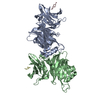
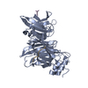
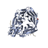
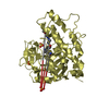
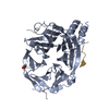
[English] 日本語
Yorodumi
- PDB-2xzg: Clathrin Terminal Domain Complexed with Pitstop 1 -

+
Open data


ID or keywords:

Loading...

-
Basic information

Entry
Database: PDB / ID: 2xzg
TitleClathrin Terminal Domain Complexed with Pitstop 1
ComponentsCLATHRIN HEAVY CHAIN 1
KeywordsENDOCYTOSIS / ENDOCYTOSIS INHIBITION
Function / homology
Function and homology information


clathrin coat of trans-Golgi network vesicle / clathrin light chain binding / negative regulation of hyaluronan biosynthetic process / clathrin complex / clathrin coat / WNT5A-dependent internalization of FZD2, FZD5 and ROR2 / WNT5A-dependent internalization of FZD4 / Entry of Influenza Virion into Host Cell via Endocytosis / transferrin transport / amyloid-beta clearance by transcytosis ...clathrin coat of trans-Golgi network vesicle / clathrin light chain binding / negative regulation of hyaluronan biosynthetic process / clathrin complex / clathrin coat / WNT5A-dependent internalization of FZD2, FZD5 and ROR2 / WNT5A-dependent internalization of FZD4 / Entry of Influenza Virion into Host Cell via Endocytosis / transferrin transport / amyloid-beta clearance by transcytosis / mitotic spindle microtubule / clathrin coat of coated pit / clathrin coat disassembly / clathrin coat assembly / Retrograde neurotrophin signalling / Formation of annular gap junctions / clathrin-coated endocytic vesicle / Gap junction degradation / LDL clearance / clathrin-dependent endocytosis / ALK mutants bind TKIs / endolysosome membrane / retrograde transport, endosome to Golgi / negative regulation of protein localization to plasma membrane / clathrin-coated vesicle / Lysosome Vesicle Biogenesis / RHOV GTPase cycle / low-density lipoprotein particle receptor binding / Golgi Associated Vesicle Biogenesis / ubiquitin-specific protease binding / Recycling pathway of L1 / RHOU GTPase cycle / EPH-ephrin mediated repulsion of cells / regulation of mitotic spindle organization / MHC class II antigen presentation / VLDLR internalisation and degradation / receptor-mediated endocytosis / trans-Golgi network membrane / intracellular protein transport / clathrin-coated endocytic vesicle membrane / receptor internalization / mitotic spindle / autophagy / spindle / osteoblast differentiation / double-stranded RNA binding / disordered domain specific binding / extracellular vesicle / Signaling by ALK fusions and activated point mutants / melanosome / Cargo recognition for clathrin-mediated endocytosis / Clathrin-mediated endocytosis / mitotic cell cycle / lysosome / endosome / cell division / focal adhesion / protein kinase binding / structural molecule activity / protein-containing complex / RNA binding / extracellular exosome / membrane / plasma membrane / cytosol
Similarity search - Function
Clathrin heavy-chain terminal domain / Clathrin, heavy chain, linker, core motif / Clathrin heavy chain, N-terminal / Clathrin, heavy chain / Clathrin, heavy chain, propeller repeat / Clathrin propeller repeat / Clathrin, heavy-chain linker / Clathrin-H-link / Region in Clathrin and VPS / Clathrin heavy chain repeat homology ...Clathrin heavy-chain terminal domain / Clathrin, heavy chain, linker, core motif / Clathrin heavy chain, N-terminal / Clathrin, heavy chain / Clathrin, heavy chain, propeller repeat / Clathrin propeller repeat / Clathrin, heavy-chain linker / Clathrin-H-link / Region in Clathrin and VPS / Clathrin heavy chain repeat homology / Clathrin, heavy chain/VPS, 7-fold repeat / Clathrin heavy-chain (CHCR) repeat profile. / 7 Propeller / Methylamine Dehydrogenase; Chain H / Tetratricopeptide-like helical domain superfamily / Armadillo-type fold / Mainly Beta
Similarity search - Domain/homology
ACETATE ION / DI(HYDROXYETHYL)ETHER / Chem-VH1 / Clathrin heavy chain 1
Similarity search - Component
Biological speciesHOMO SAPIENS (human)
MethodX-RAY DIFFRACTION / SYNCHROTRON / MIR / Resolution: 1.7 Å
AuthorsBulut, H. / Von Kleist, L. / Saenger, W. / Haucke, V.
CitationJournal: Cell(Cambridge,Mass.) / Year: 2011
Title: Role of the Clathrin Terminal Domain in Regulating Coated Pit Dynamics Revealed by Small Molecule Inhibition.
Authors: Von Kleist, L. / Stahlschmidt, W. / Bulut, H. / Gromova, K. / Puchkov, D. / Robertson, M.J. / Macgregor, K.A. / Tomlin, N. / Pechstein, A. / Chau, N. / Chircop, M. / Sakoff, J. / Von Kries, ...Authors: Von Kleist, L. / Stahlschmidt, W. / Bulut, H. / Gromova, K. / Puchkov, D. / Robertson, M.J. / Macgregor, K.A. / Tomlin, N. / Pechstein, A. / Chau, N. / Chircop, M. / Sakoff, J. / Von Kries, J.P. / Saenger, W. / Krausslich, H. / Shupliakov, O. / Robinson, P.J. / Mccluskey, A. / Haucke, V.
History
DepositionNov 25, 2010Deposition site: PDBE / Processing site: PDBE
Revision 1.0Aug 17, 2011Provider: repository / Type: Initial release
Revision 1.1Dec 20, 2023Group: Data collection / Database references ...Data collection / Database references / Derived calculations / Other / Refinement description
Category: chem_comp_atom / chem_comp_bond ...chem_comp_atom / chem_comp_bond / database_2 / pdbx_database_status / pdbx_initial_refinement_model / struct_site
Item: _database_2.pdbx_DOI / _database_2.pdbx_database_accession ..._database_2.pdbx_DOI / _database_2.pdbx_database_accession / _pdbx_database_status.status_code_sf / _struct_site.pdbx_auth_asym_id / _struct_site.pdbx_auth_comp_id / _struct_site.pdbx_auth_seq_id

-
Structure visualization

Structure viewerMolecule:
MolmilJmol/JSmol

Downloads & links

-
Assembly

Deposited unit
A: CLATHRIN HEAVY CHAIN 1
hetero molecules


Theoretical massNumber of molelcules
Total (without water)41,4366
Polymers40,6911
Non-polymers7465
Water8,251458
1


  • Idetical with deposited unit
  • defined by author&software
TypeNameSymmetry operationNumber
identity operation1_555x,y,z1
MethodPISA
Unit cell
Length a, b, c (Å)71.340, 73.915, 84.384
Angle α, β, γ (deg.)90.00, 90.00, 90.00
Int Tables number19
Space group name H-MP212121

-
Components

-
Antibody , 1 types, 1 molecules A

#1: Antibody CLATHRIN HEAVY CHAIN 1 / CLATHRIN HEAVY CHAIN ON CHROMOSOME 17 / CLH-17


Mass: 40690.688 Da / Num. of mol.: 1 / Fragment: N-TERMINAL DOMAIN, RESIDUES 1-364
Source method: isolated from a genetically manipulated source
Source: (gene. exp.) HOMO SAPIENS (human) / Production host: ESCHERICHIA COLI (E. coli) / References: UniProt: Q00610

-
Non-polymers , 5 types, 463 molecules

#2: Chemical ChemComp-PEG / DI(HYDROXYETHYL)ETHER


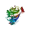
Mass: 106.120 Da / Num. of mol.: 2 / Source method: obtained synthetically / Formula: C4H10O3
#3: Chemical ChemComp-GOL / GLYCEROL / GLYCERIN / PROPANE-1,2,3-TRIOL


Mass: 92.094 Da / Num. of mol.: 1 / Source method: obtained synthetically / Formula: C3H8O3
#4: Chemical ChemComp-ACT / ACETATE ION


Mass: 59.044 Da / Num. of mol.: 1 / Source method: obtained synthetically / Formula: C2H3O2
#5: Chemical ChemComp-VH1 / 2-(4-AMINOBENZYL)-1,3-DIOXO-2,3-DIHYDRO-1H-BENZO[DE]ISOQUINOLINE-5-SULFONATE


Mass: 382.390 Da / Num. of mol.: 1 / Source method: obtained synthetically / Formula: C19H13N2O5S
#6: Water ChemComp-HOH / water


Mass: 18.015 Da / Num. of mol.: 458 / Source method: isolated from a natural source / Formula: H2O

-
Details

Nonpolymer detailsPITSTOP 1 COMPOUND IS RESIDUE VH1, 2-(4-AMINOBENZYL)- 1,3-DIOXO-2,3-DIHYDRO-1H-BENZO[DE] ...PITSTOP 1 COMPOUND IS RESIDUE VH1, 2-(4-AMINOBENZYL)- 1,3-DIOXO-2,3-DIHYDRO-1H-BENZO[DE]ISOQUINOLINE-5-SULFONATE.

-
Experimental details

-
Experiment

ExperimentMethod: X-RAY DIFFRACTION / Number of used crystals: 1

-
Sample preparation

CrystalDensity Matthews: 2.72 Å3/Da / Density % sol: 54.75 % / Description: NONE
Crystal growpH: 8
Details: 20% PEG 3350, 150 MM POTASSIUM ACETATE, 0.1 M TRIS, PH 8.0.

-
Data collection

DiffractionMean temperature: 291 K
Diffraction sourceSource: SYNCHROTRON / Site: BESSY / Beamline: 14.1 / Wavelength: 0.91841
DetectorType: MARMOSAIC 225 mm CCD / Detector: CCD / Date: Oct 7, 2009
RadiationProtocol: SINGLE WAVELENGTH / Monochromatic (M) / Laue (L): M / Scattering type: x-ray
Radiation wavelengthWavelength: 0.91841 Å / Relative weight: 1
ReflectionResolution: 1.7→33.85 Å / Num. obs: 47297 / % possible obs: 95.1 % / Observed criterion σ(I): -3 / Redundancy: 5.9 % / Rmerge(I) obs: 0.05 / Net I/σ(I): 24.34
Reflection shellHighest resolution: 1.7 Å / Redundancy: 2.6 % / Rmerge(I) obs: 0.24 / Mean I/σ(I) obs: 4.19 / % possible all: 1.7

-
Processing

Software
NameVersionClassification
REFMAC5.5.0110refinement
XDSdata reduction
XDSdata scaling
MOLREPphasing
RefinementMethod to determine structure: MIR
Starting model: PDB ENTRY 1C9I
Resolution: 1.7→33.85 Å / Cor.coef. Fo:Fc: 0.962 / Cor.coef. Fo:Fc free: 0.942 / SU B: 1.679 / SU ML: 0.056 / Cross valid method: THROUGHOUT / ESU R: 0.089 / ESU R Free: 0.094 / Stereochemistry target values: MAXIMUM LIKELIHOOD / Details: HYDROGENS HAVE BEEN ADDED IN THE RIDING POSITIONS.
RfactorNum. reflection% reflectionSelection details
Rfree0.19973 2366 5 %RANDOM
Rwork0.15864 ---
obs0.16071 44946 100 %-
Solvent computationIon probe radii: 0.8 Å / Shrinkage radii: 0.8 Å / VDW probe radii: 1.4 Å / Solvent model: MASK
Displacement parametersBiso mean: 12.556 Å2
Baniso -1Baniso -2Baniso -3
1--0.48 Å20 Å20 Å2
2---0.8 Å20 Å2
3---1.28 Å2
Refinement stepCycle: LAST / Resolution: 1.7→33.85 Å
ProteinNucleic acidLigandSolventTotal
Num. atoms2803 0 51 458 3312
Refine LS restraints
Refine-IDTypeDev idealDev ideal targetNumber
X-RAY DIFFRACTIONr_bond_refined_d0.0230.0222945
X-RAY DIFFRACTIONr_bond_other_d0.0010.021971
X-RAY DIFFRACTIONr_angle_refined_deg1.8651.9633996
X-RAY DIFFRACTIONr_angle_other_deg1.0063.0014825
X-RAY DIFFRACTIONr_dihedral_angle_1_deg6.6515365
X-RAY DIFFRACTIONr_dihedral_angle_2_deg36.49825128
X-RAY DIFFRACTIONr_dihedral_angle_3_deg13.39815507
X-RAY DIFFRACTIONr_dihedral_angle_4_deg19.2981513
X-RAY DIFFRACTIONr_chiral_restr0.1220.2448
X-RAY DIFFRACTIONr_gen_planes_refined0.0090.0213252
X-RAY DIFFRACTIONr_gen_planes_other0.0010.02554
X-RAY DIFFRACTIONr_nbd_refined
X-RAY DIFFRACTIONr_nbd_other
X-RAY DIFFRACTIONr_nbtor_refined
X-RAY DIFFRACTIONr_nbtor_other
X-RAY DIFFRACTIONr_xyhbond_nbd_refined
X-RAY DIFFRACTIONr_xyhbond_nbd_other
X-RAY DIFFRACTIONr_metal_ion_refined
X-RAY DIFFRACTIONr_metal_ion_other
X-RAY DIFFRACTIONr_symmetry_vdw_refined
X-RAY DIFFRACTIONr_symmetry_vdw_other
X-RAY DIFFRACTIONr_symmetry_hbond_refined
X-RAY DIFFRACTIONr_symmetry_hbond_other
X-RAY DIFFRACTIONr_symmetry_metal_ion_refined
X-RAY DIFFRACTIONr_symmetry_metal_ion_other
X-RAY DIFFRACTIONr_mcbond_it1.2751.51813
X-RAY DIFFRACTIONr_mcbond_other0.3741.5726
X-RAY DIFFRACTIONr_mcangle_it2.11622951
X-RAY DIFFRACTIONr_mcangle_other
X-RAY DIFFRACTIONr_scbond_it2.97731132
X-RAY DIFFRACTIONr_scbond_other
X-RAY DIFFRACTIONr_scangle_it4.6634.51045
X-RAY DIFFRACTIONr_scangle_other
X-RAY DIFFRACTIONr_long_range_B_refined
X-RAY DIFFRACTIONr_long_range_B_other
X-RAY DIFFRACTIONr_rigid_bond_restr
X-RAY DIFFRACTIONr_sphericity_free
X-RAY DIFFRACTIONr_sphericity_bonded
LS refinement shellResolution: 1.7→1.744 Å / Total num. of bins used: 20
RfactorNum. reflection% reflection
Rfree0.283 126 -
Rwork0.221 2404 -
obs--100 %

+
About Yorodumi

-
News

-
Feb 9, 2022. New format data for meta-information of EMDB entries

New format data for meta-information of EMDB entries

  • Version 3 of the EMDB header file is now the official format.
  • The previous official version 1.9 will be removed from the archive.

Related info.:EMDB header

External links:wwPDB to switch to version 3 of the EMDB data model

-
Aug 12, 2020. Covid-19 info

Covid-19 info

URL: https://pdbjlvh1.pdbj.org/emnavi/covid19.php

New page: Covid-19 featured information page in EM Navigator.

Related info.:Covid-19 info / Mar 5, 2020. Novel coronavirus structure data

+
Mar 5, 2020. Novel coronavirus structure data

Novel coronavirus structure data

Related info.:Yorodumi Speices / Aug 12, 2020. Covid-19 info

External links:COVID-19 featured content - PDBj / Molecule of the Month (242):Coronavirus Proteases

+
Jan 31, 2019. EMDB accession codes are about to change! (news from PDBe EMDB page)

EMDB accession codes are about to change! (news from PDBe EMDB page)

  • The allocation of 4 digits for EMDB accession codes will soon come to an end. Whilst these codes will remain in use, new EMDB accession codes will include an additional digit and will expand incrementally as the available range of codes is exhausted. The current 4-digit format prefixed with “EMD-” (i.e. EMD-XXXX) will advance to a 5-digit format (i.e. EMD-XXXXX), and so on. It is currently estimated that the 4-digit codes will be depleted around Spring 2019, at which point the 5-digit format will come into force.
  • The EM Navigator/Yorodumi systems omit the EMD- prefix.

Related info.:Q: What is EMD? / ID/Accession-code notation in Yorodumi/EM Navigator

External links:EMDB Accession Codes are Changing Soon! / Contact to PDBj

+
Jul 12, 2017. Major update of PDB

Major update of PDB

  • wwPDB released updated PDB data conforming to the new PDBx/mmCIF dictionary.
  • This is a major update changing the version number from 4 to 5, and with Remediation, in which all the entries are updated.
  • In this update, many items about electron microscopy experimental information are reorganized (e.g. em_software).
  • Now, EM Navigator and Yorodumi are based on the updated data.

External links:wwPDB Remediation / Enriched Model Files Conforming to OneDep Data Standards Now Available in the PDB FTP Archive

-
Yorodumi

Thousand views of thousand structures

  • Yorodumi is a browser for structure data from EMDB, PDB, SASBDB, etc.
  • This page is also the successor to EM Navigator detail page, and also detail information page/front-end page for Omokage search.
  • The word "yorodu" (or yorozu) is an old Japanese word meaning "ten thousand". "mi" (miru) is to see.

Related info.:EMDB / PDB / SASBDB / Comparison of 3 databanks / Yorodumi Search / Aug 31, 2016. New EM Navigator & Yorodumi / Yorodumi Papers / Jmol/JSmol / Function and homology information / Changes in new EM Navigator and Yorodumi

Read more