 Movie
Movie Controller

[English] 日本語
 Yorodumi
- PDB-1c9i: PEPTIDE-IN-GROOVE INTERACTIONS LINK TARGET PROTEINS TO THE B-PROP... -
+ Open data

- Basic information
| Entry | Database: PDB / ID: 1c9i | ||||||
|---|---|---|---|---|---|---|---|
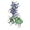
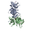
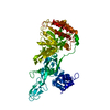
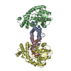
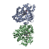
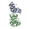
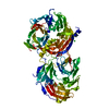
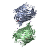
| Title | PEPTIDE-IN-GROOVE INTERACTIONS LINK TARGET PROTEINS TO THE B-PROPELLER OF CLATHRIN | ||||||
 Components Components |
| ||||||
 Keywords Keywords | ENDOCYTOSIS/EXOCYTOSIS / BETA-PROPELLER / HELICAL HAIRPIN / ENDOCYTOSIS-EXOCYTOSIS COMPLEX | ||||||
| Function / homology |  Function and homology information Function and homology informationpresynaptic endocytic zone membrane / RHOU GTPase cycle / RHOV GTPase cycle / clathrin coat of trans-Golgi network vesicle / Myb complex / Gap junction degradation / Formation of annular gap junctions / clathrin light chain binding / WNT5A-dependent internalization of FZD2, FZD5 and ROR2 / LDL clearance ...presynaptic endocytic zone membrane / RHOU GTPase cycle / RHOV GTPase cycle / clathrin coat of trans-Golgi network vesicle / Myb complex / Gap junction degradation / Formation of annular gap junctions / clathrin light chain binding / WNT5A-dependent internalization of FZD2, FZD5 and ROR2 / LDL clearance / negative regulation of hyaluronan biosynthetic process / Retrograde neurotrophin signalling / VLDLR internalisation and degradation / clathrin complex / Lysosome Vesicle Biogenesis / WNT5A-dependent internalization of FZD4 / clathrin coat / MHC class II antigen presentation / amyloid-beta clearance by transcytosis / clathrin coat of coated pit / Golgi Associated Vesicle Biogenesis / photoreceptor ribbon synapse / postsynaptic endocytic zone / clathrin coat disassembly / extrinsic component of synaptic vesicle membrane / Recycling pathway of L1 / clathrin-coated endocytic vesicle / membrane coat / Cargo recognition for clathrin-mediated endocytosis / clathrin coat assembly / mitotic spindle microtubule / Clathrin-mediated endocytosis / clathrin-dependent endocytosis / retrograde transport, endosome to Golgi / clathrin-coated vesicle / ankyrin binding / low-density lipoprotein particle receptor binding / ubiquitin-specific protease binding / Golgi organization / mitotic spindle assembly / negative regulation of protein localization to plasma membrane / synaptic vesicle endocytosis / heat shock protein binding / peptide binding / clathrin-coated pit / T-tubule / receptor-mediated endocytosis / regulation of mitotic spindle organization / protein serine/threonine kinase binding / clathrin-coated endocytic vesicle membrane / transferrin transport / intracellular protein transport / sarcolemma / receptor internalization / spindle / autophagy / centriolar satellite / disordered domain specific binding / terminal bouton / mitotic spindle / melanosome / mitotic cell cycle / double-stranded RNA binding / lysosome / endosome / protein kinase binding / structural molecule activity / glutamatergic synapse / protein-containing complex / membrane / cytosol Similarity search - Function | ||||||
| Biological species |  | ||||||
| Method |  X-RAY DIFFRACTION / Resolution: 2.9 Å X-RAY DIFFRACTION / Resolution: 2.9 Å | ||||||
 Authors Authors | ter Haar, E. / Harrison, S.C. / Kirchhausen, T. | ||||||
 Citation Citation | Journal: Proc.Natl.Acad.Sci.USA / Year: 2000 Title: Peptide-in-groove interactions link target proteins to the beta-propeller of clathrin. Authors: ter Haar, E. / Harrison, S.C. / Kirchhausen, T. | ||||||
| History |
|
- Structure visualization
| Structure viewer | Molecule: MolmilJmol/JSmol |
|---|
- Downloads & links
-Download
| PDBx/mmCIF format | 1c9i.cif.gz | 145.6 KB | Display | PDBx/mmCIF format |
|---|---|---|---|---|
| PDB format | pdb1c9i.ent.gz | 117.1 KB | Display | PDB format |
| PDBx/mmJSON format | 1c9i.json.gz | Tree view | PDBx/mmJSON format | |
| Others |  Other downloads Other downloads |
-Validation report
| Arichive directory | https://data.pdbj.org/pub/pdb/validation_reports/c9/1c9iftp://data.pdbj.org/pub/pdb/validation_reports/c9/1c9i | HTTPS FTP |
|---|
-Related structure data


-Links
 PDBj
PDBj


- Assembly
| Deposited unit | 
| ||||||||
|---|---|---|---|---|---|---|---|---|---|
| 1 |
| ||||||||
| Unit cell |
|
-Components
| #1: Protein | Mass: 39953.879 Da / Num. of mol.: 2 / Fragment: N-TERMINAL DOMAIN Source method: isolated from a genetically manipulated source Source: (gene. exp.)  #2: Protein/peptide | Mass: 916.028 Da / Num. of mol.: 2 / Fragment: CLATHRIN-BOX PEPTIDE / Mutation: D815A, D823A / Source method: obtained synthetically Details: This peptide was chemically sythesized.The sequence of this peptide naturally occurs in humans (HOMO SAPIENS). |
|---|
-Experimental details
-Experiment
| Experiment | Method: X-RAY DIFFRACTION / Number of used crystals: 1 |
|---|
- Sample preparation
| Crystal | Density Matthews: 3.9 Å3/Da / Density % sol: 68.42 % | |||||||||||||||||||||||||||||||||||
|---|---|---|---|---|---|---|---|---|---|---|---|---|---|---|---|---|---|---|---|---|---|---|---|---|---|---|---|---|---|---|---|---|---|---|---|---|
| Crystal grow | Temperature: 298 K / Method: vapor diffusion, hanging drop / pH: 8 Details: PEG 4000, KOAc, DTT, Tris, pH 8.0, VAPOR DIFFUSION, HANGING DROP, temperature 298K | |||||||||||||||||||||||||||||||||||
| Crystal grow | *PLUS | |||||||||||||||||||||||||||||||||||
| Components of the solutions | *PLUS
|
-Data collection
| Diffraction | Mean temperature: 100 K |
|---|---|
| Diffraction source | Source: ROTATING ANODE / Type: ELLIOTT GX-13 / Wavelength: 1.54 |
| Detector | Type: MARRESEARCH / Detector: IMAGE PLATE / Date: Apr 1, 1999 |
| Radiation | Protocol: SINGLE WAVELENGTH / Monochromatic (M) / Laue (L): M / Scattering type: x-ray |
| Radiation wavelength | Wavelength: 1.54 Å / Relative weight: 1 |
| Reflection | Resolution: 2.7→30 Å / Num. all: 27038 / Num. obs: 27038 / % possible obs: 97.3 % / Observed criterion σ(F): 0 / Observed criterion σ(I): 0 / Redundancy: 7.1 % / Biso Wilson estimate: 44.7 Å2 / Rmerge(I) obs: 0.096 / Net I/σ(I): 12.3 |
| Reflection shell | Resolution: 2.9→3.08 Å / Redundancy: 3.2 % / Rmerge(I) obs: 0.207 / % possible all: 94.6 |
| Reflection shell | *PLUS % possible obs: 94.6 % |
- Processing
| Software |
| |||||||||||||||||||||||||
|---|---|---|---|---|---|---|---|---|---|---|---|---|---|---|---|---|---|---|---|---|---|---|---|---|---|---|
| Refinement | Resolution: 2.9→30 Å / Rfactor Rfree error: 0.008 / Data cutoff high absF: 277796.29 / Data cutoff low absF: 0 / Isotropic thermal model: RESTRAINED / Cross valid method: THROUGHOUT / σ(F): 0 / σ(I): 0 / Stereochemistry target values: Engh & Huber
| |||||||||||||||||||||||||
| Solvent computation | Solvent model: FLAT MODEL / Bsol: 29.83 Å2 / ksol: 0.387 e/Å3 | |||||||||||||||||||||||||
| Displacement parameters | Biso mean: 19.9 Å2
| |||||||||||||||||||||||||
| Refine analyze |
| |||||||||||||||||||||||||
| Refinement step | Cycle: LAST / Resolution: 2.9→30 Å
| |||||||||||||||||||||||||
| Refine LS restraints |
| |||||||||||||||||||||||||
| LS refinement shell | Resolution: 2.9→3.08 Å / Rfactor Rfree error: 0.023 / Total num. of bins used: 6
| |||||||||||||||||||||||||
| Xplor file | Serial no: 1 / Param file: PROTEIN_REP.PA / Topol file: PROTEIN.TOP | |||||||||||||||||||||||||
| Software | *PLUS Name: CNS / Version: 0.5 / Classification: refinement | |||||||||||||||||||||||||
| Refinement | *PLUS Rfactor Rfree: 0.268 | |||||||||||||||||||||||||
| Solvent computation | *PLUS | |||||||||||||||||||||||||
| Displacement parameters | *PLUS |
