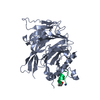
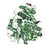
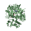
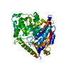
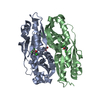
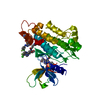
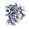
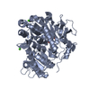
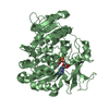
Entry Database : PDB / ID : 2xyiTitle Crystal Structure of Nurf55 in complex with a H4 peptide HISTONE H4 PROBABLE HISTONE-BINDING PROTEIN CAF1 Keywords / / / Function / homology Function Domain/homology Component
/ / / / / / / / / / / / / / / / / / / / / / / / / / / / / / / / / / / / / / / / / / / / / / / / / / / / / / / / / / / / / / / / / / / / / / / / / / / / / / / / / / / / / / / / / / / / / / / / / / / / / / / / / / Biological species DROSOPHILA MELANOGASTER (fruit fly)Method / / / Resolution : 1.75 Å Authors Stirnimann, C.U. / Nowak, A.J. / Mueller, C.W. Journal : J.Biol.Chem. / Year : 2011Title : Chromatin-Modifying Complex Component Nurf55/P55 Associates with Histones H3, H4 and Polycomb Repressive Complex 2 Subunit Su(Z)12 Through Partially Overlapping Binding Sites.Authors : Nowak, A.J. / Alfieri, C. / Stirnimann, C.U. / Rybin, V. / Baudin, F. / Ly-Hartig, N. / Lindner, D. / Muller, C.W. History Deposition Nov 17, 2010 Deposition site / Processing site Revision 1.0 May 4, 2011 Provider / Type Revision 1.1 Jun 30, 2011 Group Revision 1.2 Jul 13, 2011 Group Revision 1.3 Apr 24, 2019 Group Advisory / Data collection ... Advisory / Data collection / Other / Source and taxonomy Category entity_src_gen / pdbx_database_proc ... entity_src_gen / pdbx_database_proc / pdbx_database_status / pdbx_unobs_or_zero_occ_atoms Item / _pdbx_database_status.recvd_author_approvalRevision 1.4 Oct 9, 2019 Group / Experimental preparation / OtherCategory / pdbx_database_status / reflnsItem / _pdbx_database_status.status_code_sf / _reflns.pdbx_Rmerge_I_obsRevision 1.5 Dec 20, 2023 Group Advisory / Data collection ... Advisory / Data collection / Database references / Derived calculations / Refinement description Category chem_comp_atom / chem_comp_bond ... chem_comp_atom / chem_comp_bond / database_2 / pdbx_initial_refinement_model / pdbx_unobs_or_zero_occ_atoms / struct_site Item _database_2.pdbx_DOI / _database_2.pdbx_database_accession ... _database_2.pdbx_DOI / _database_2.pdbx_database_accession / _struct_site.pdbx_auth_asym_id / _struct_site.pdbx_auth_comp_id / _struct_site.pdbx_auth_seq_id
Show all Show less
 Movie
Movie Controller


 Open data
 Basic information
 Components
 Keywords
 Function and homology information
 X-RAY DIFFRACTION /
  Authors
 Citation
 Structure visualization
 Downloads & links
 Other downloads
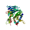

 PDBj
PDBj


 Assembly
 SPODOPTERA FRUGIPERDA (fall armyworm) / References: UniProt: Q24572
 Mass: 194.226 Da / Num. of mol.: 3 / Source method: obtained synthetically / Formula: C8H18O5 / Comment: precipitant*YM
 Mass: 106.120 Da / Num. of mol.: 2 / Source method: obtained synthetically / Formula: C4H10O3
 Mass: 18.015 Da / Num. of mol.: 398 / Source method: isolated from a natural source / Formula: H2O
 Sample preparation
 / Beamline: ID23-2 / Wavelength: 1.0085
  Processing