[English] 日本語
Yorodumi
- PDB-3o84: Structure of BasE N-terminal domain from Acinetobacter baumannii ... -

+
Open data


ID or keywords:

Loading...

-
Basic information

Entry
Database: PDB / ID: 3o84
Title
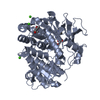
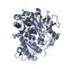
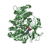
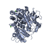
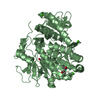
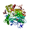
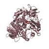
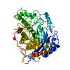
Structure of BasE N-terminal domain from Acinetobacter baumannii bound to 6-phenyl-1-(pyridin-4-ylmethyl)-1H-pyrazolo[3,4-b]pyridine-4-carboxylic acid.
ComponentsPeptide arylation enzyme
KeywordsLIGASE / Adenylation of 2 / 3-dihydroxybenzoate and transfer to pantetheine cofactor of BasF / Non-Ribosomal Peptide Synthetase (NRPS)
Function / homology
Function and homology information


2,3-dihydroxybenzoate--[aryl-carrier protein] ligase / siderophore biosynthetic process
Similarity search - Function
2,3-dihydroxybenzoate-AMP ligase / ANL, N-terminal domain / ANL, N-terminal domain / AMP-binding enzyme, C-terminal domain / AMP-binding enzyme C-terminal domain / AMP-binding, conserved site / Putative AMP-binding domain signature. / AMP-dependent synthetase/ligase / AMP-binding enzyme, C-terminal domain superfamily / AMP-binding enzyme ...2,3-dihydroxybenzoate-AMP ligase / ANL, N-terminal domain / ANL, N-terminal domain / AMP-binding enzyme, C-terminal domain / AMP-binding enzyme C-terminal domain / AMP-binding, conserved site / Putative AMP-binding domain signature. / AMP-dependent synthetase/ligase / AMP-binding enzyme, C-terminal domain superfamily / AMP-binding enzyme / Rossmann fold / 3-Layer(aba) Sandwich / Alpha Beta
Similarity search - Domain/homology
Chem-HTJ / TRIETHYLENE GLYCOL / Peptide arylation enzyme / Peptide arylation enzyme
Similarity search - Component
Biological speciesAcinetobacter baumannii (bacteria)
MethodX-RAY DIFFRACTION / SYNCHROTRON / FOURIER SYNTHESIS / Resolution: 2.1 Å
AuthorsDrake, E.J. / Duckworth, B.P. / Neres, J. / Aldrich, C.C. / Gulick, A.M.
CitationJournal: Biochemistry / Year: 2010
Title: Biochemical and structural characterization of bisubstrate inhibitors of BasE, the self-standing nonribosomal peptide synthetase adenylate-forming enzyme of acinetobactin synthesis.
Authors: Drake, E.J. / Duckworth, B.P. / Neres, J. / Aldrich, C.C. / Gulick, A.M.
History
DepositionAug 2, 2010Deposition site: RCSB / Processing site: RCSB
Revision 1.0Oct 6, 2010Provider: repository / Type: Initial release
Revision 1.1Jul 13, 2011Group: Version format compliance
Revision 1.2Nov 8, 2017Group: Refinement description / Category: software / Item: _software.classification / _software.name
Revision 1.3Sep 6, 2023Group: Data collection / Database references ...Data collection / Database references / Derived calculations / Refinement description
Category: chem_comp_atom / chem_comp_bond ...chem_comp_atom / chem_comp_bond / database_2 / pdbx_initial_refinement_model / pdbx_struct_conn_angle / struct_conn / struct_ref_seq_dif / struct_site
Item: _database_2.pdbx_DOI / _database_2.pdbx_database_accession ..._database_2.pdbx_DOI / _database_2.pdbx_database_accession / _pdbx_struct_conn_angle.ptnr1_auth_asym_id / _pdbx_struct_conn_angle.ptnr1_auth_comp_id / _pdbx_struct_conn_angle.ptnr1_auth_seq_id / _pdbx_struct_conn_angle.ptnr1_label_asym_id / _pdbx_struct_conn_angle.ptnr1_label_atom_id / _pdbx_struct_conn_angle.ptnr1_label_comp_id / _pdbx_struct_conn_angle.ptnr1_label_seq_id / _pdbx_struct_conn_angle.ptnr2_auth_asym_id / _pdbx_struct_conn_angle.ptnr2_auth_seq_id / _pdbx_struct_conn_angle.ptnr2_label_asym_id / _pdbx_struct_conn_angle.ptnr3_auth_asym_id / _pdbx_struct_conn_angle.ptnr3_auth_comp_id / _pdbx_struct_conn_angle.ptnr3_auth_seq_id / _pdbx_struct_conn_angle.ptnr3_label_asym_id / _pdbx_struct_conn_angle.ptnr3_label_atom_id / _pdbx_struct_conn_angle.ptnr3_label_comp_id / _pdbx_struct_conn_angle.ptnr3_label_seq_id / _pdbx_struct_conn_angle.value / _struct_conn.pdbx_dist_value / _struct_conn.ptnr1_auth_asym_id / _struct_conn.ptnr1_auth_comp_id / _struct_conn.ptnr1_auth_seq_id / _struct_conn.ptnr1_label_asym_id / _struct_conn.ptnr1_label_atom_id / _struct_conn.ptnr1_label_comp_id / _struct_conn.ptnr1_label_seq_id / _struct_conn.ptnr2_auth_asym_id / _struct_conn.ptnr2_auth_comp_id / _struct_conn.ptnr2_auth_seq_id / _struct_conn.ptnr2_label_asym_id / _struct_conn.ptnr2_label_atom_id / _struct_conn.ptnr2_label_comp_id / _struct_ref_seq_dif.details / _struct_site.pdbx_auth_asym_id / _struct_site.pdbx_auth_comp_id / _struct_site.pdbx_auth_seq_id

-
Structure visualization

Structure viewerMolecule:
MolmilJmol/JSmol

Downloads & links

-
Assembly

Deposited unit
A: Peptide arylation enzyme
B: Peptide arylation enzyme
hetero molecules


Theoretical massNumber of molelcules
Total (without water)122,94411
Polymers121,7372
Non-polymers1,2089
Water7,819434
1
A: Peptide arylation enzyme
hetero molecules


Theoretical massNumber of molelcules
Total (without water)61,4376
Polymers60,8681
Non-polymers5695
Water181
TypeNameSymmetry operationNumber
identity operation1_555x,y,z1
2
B: Peptide arylation enzyme
hetero molecules


Theoretical massNumber of molelcules
Total (without water)61,5075
Polymers60,8681
Non-polymers6394
Water181
TypeNameSymmetry operationNumber
identity operation1_555x,y,z1
3


  • Idetical with deposited unit
  • defined by software
TypeNameSymmetry operationNumber
identity operation1_555x,y,z1
Buried area2550 Å2
ΔGint-55 kcal/mol
Surface area34110 Å2
MethodPISA
Unit cell
Length a, b, c (Å)65.534, 143.312, 148.815
Angle α, β, γ (deg.)90.00, 90.00, 90.00
Int Tables number19
Space group name H-MP212121

-
Components

-
Protein , 1 types, 2 molecules AB

#1: Protein Peptide arylation enzyme


Mass: 60868.270 Da / Num. of mol.: 2 / Fragment: BasE / Mutation: P45L
Source method: isolated from a genetically manipulated source
Source: (gene. exp.) Acinetobacter baumannii (bacteria) / Strain: AB900 / Gene: ACICU_02578, basE / Plasmid: pED453 / Production host: Escherichia coli (E. coli) / Strain (production host): BL21(DE3)
References: UniProt: B2HVG8, UniProt: A0A7U3Y1M5*PLUS, Ligases; Forming carbon-sulfur bonds; Acid-thiol ligases

-
Non-polymers , 6 types, 443 molecules

#2: Chemical ChemComp-HTJ / 6-phenyl-1-(pyridin-4-ylmethyl)-1H-pyrazolo[3,4-b]pyridine-4-carboxylic acid


Mass: 330.340 Da / Num. of mol.: 2 / Source method: obtained synthetically / Formula: C19H14N4O2
#3: Chemical
ChemComp-CA / CALCIUM ION


Mass: 40.078 Da / Num. of mol.: 4 / Source method: obtained synthetically / Formula: Ca
#4: Chemical ChemComp-MPD / (4S)-2-METHYL-2,4-PENTANEDIOL / 2-Methyl-2,4-pentanediol


Mass: 118.174 Da / Num. of mol.: 1 / Source method: obtained synthetically / Formula: C6H14O2 / Comment: precipitant*YM
#5: Chemical ChemComp-MRD / (4R)-2-METHYLPENTANE-2,4-DIOL / 2-Methyl-2,4-pentanediol


Mass: 118.174 Da / Num. of mol.: 1 / Source method: obtained synthetically / Formula: C6H14O2 / Comment: precipitant*YM
#6: Chemical ChemComp-PGE / TRIETHYLENE GLYCOL / Polyethylene glycol


Mass: 150.173 Da / Num. of mol.: 1 / Source method: obtained synthetically / Formula: C6H14O4
#7: Water ChemComp-HOH / water / Water


Mass: 18.015 Da / Num. of mol.: 434 / Source method: isolated from a natural source / Formula: H2O

-
Details

Sequence detailsTHIS ENTRY USES A UNIPROT REFERENCE THAT IS FOR A DIFFERENT STRAIN OF A. BAUMANNI. THESE CHANGES ...THIS ENTRY USES A UNIPROT REFERENCE THAT IS FOR A DIFFERENT STRAIN OF A. BAUMANNI. THESE CHANGES ARE STRAIN RELATED DIFFERENCES AS FOUND IN GENBANK ENTRY ZP_04661818.

-
Experimental details

-
Experiment

ExperimentMethod: X-RAY DIFFRACTION / Number of used crystals: 1

-
Sample preparation

CrystalDensity Matthews: 2.87 Å3/Da / Density % sol: 57.15 %
Crystal growTemperature: 287 K / Method: vapor diffusion, hanging drop / pH: 7.5
Details: 5-15% PEG 8000, 5% MPD, 250-600 mM CaCl2, 50 mM BTP , pH 7.5, VAPOR DIFFUSION, HANGING DROP, temperature 287K

-
Data collection

DiffractionMean temperature: 113 K
Diffraction sourceSource: SYNCHROTRON / Site: CHESS / Beamline: A1 / Wavelength: 0.9782 Å
DetectorType: ADSC QUANTUM 210 / Detector: CCD / Date: Apr 24, 2009
RadiationMonochromator: horizontal focusing / Protocol: SINGLE WAVELENGTH / Monochromatic (M) / Laue (L): M / Scattering type: x-ray
Radiation wavelengthWavelength: 0.9782 Å / Relative weight: 1
ReflectionResolution: 2.1→30 Å / Num. all: 82723 / Num. obs: 82475 / % possible obs: 99.7 % / Observed criterion σ(F): 0 / Observed criterion σ(I): -3 / Redundancy: 4 % / Biso Wilson estimate: 32.3 Å2 / Rsym value: 0.072 / Net I/σ(I): 10.8
Reflection shellResolution: 2.1→2.2 Å / Redundancy: 3.34 % / Mean I/σ(I) obs: 1.8 / Num. unique all: 10447 / Rsym value: 0.425 / % possible all: 97.9

-
Processing

Software
NameVersionClassification
AdxvPackagedata processing
MOLREPphasing
REFMAC5.5.0088refinement
HKL-2000data reduction
HKL-2000data scaling
RefinementMethod to determine structure: FOURIER SYNTHESIS
Starting model: PDB ENTRY 3O83
Resolution: 2.1→29.8 Å / Cor.coef. Fo:Fc: 0.959 / Cor.coef. Fo:Fc free: 0.947 / SU B: 3.567 / SU ML: 0.095 / Cross valid method: THROUGHOUT / ESU R: 0.153 / ESU R Free: 0.138 / Stereochemistry target values: MAXIMUM LIKELIHOOD / Details: HYDROGENS HAVE BEEN ADDED IN THE RIDING POSITIONS
RfactorNum. reflection% reflectionSelection details
Rfree0.2107 4086 5 %RANDOM
Rwork0.18521 ---
all0.1865 78200 --
obs0.1865 78200 99.7 %-
Solvent computationIon probe radii: 0.8 Å / Shrinkage radii: 0.8 Å / VDW probe radii: 1.4 Å / Solvent model: MASK
Displacement parametersBiso mean: 34.368 Å2
Baniso -1Baniso -2Baniso -3
1-1.41 Å2-0 Å2-0 Å2
2--0.67 Å20 Å2
3----2.07 Å2
Refinement stepCycle: LAST / Resolution: 2.1→29.8 Å
ProteinNucleic acidLigandSolventTotal
Num. atoms6778 0 80 434 7292
Refine LS restraints
Refine-IDTypeDev idealDev ideal targetNumber
X-RAY DIFFRACTIONr_bond_refined_d0.010.0227027
X-RAY DIFFRACTIONr_angle_refined_deg1.1821.979570
X-RAY DIFFRACTIONr_dihedral_angle_1_deg5.7055872
X-RAY DIFFRACTIONr_dihedral_angle_2_deg39.43624.465327
X-RAY DIFFRACTIONr_dihedral_angle_3_deg13.834151114
X-RAY DIFFRACTIONr_dihedral_angle_4_deg17.1391539
X-RAY DIFFRACTIONr_chiral_restr0.0780.21065
X-RAY DIFFRACTIONr_gen_planes_refined0.0050.0215419
X-RAY DIFFRACTIONr_mcbond_it0.6241.54342
X-RAY DIFFRACTIONr_mcangle_it1.19627012
X-RAY DIFFRACTIONr_scbond_it1.73532685
X-RAY DIFFRACTIONr_scangle_it3.0024.52555
LS refinement shellResolution: 2.1→2.154 Å / Total num. of bins used: 20
RfactorNum. reflection% reflection
Rfree0.314 274 -
Rwork0.265 5523 -
obs--96.97 %

+
About Yorodumi

-
News

-
Feb 9, 2022. New format data for meta-information of EMDB entries

New format data for meta-information of EMDB entries

  • Version 3 of the EMDB header file is now the official format.
  • The previous official version 1.9 will be removed from the archive.

Related info.:EMDB header

External links:wwPDB to switch to version 3 of the EMDB data model

-
Aug 12, 2020. Covid-19 info

Covid-19 info

URL: https://pdbj.org/emnavi/covid19.php

New page: Covid-19 featured information page in EM Navigator.

Related info.:Covid-19 info / Mar 5, 2020. Novel coronavirus structure data

+
Mar 5, 2020. Novel coronavirus structure data

Novel coronavirus structure data

Related info.:Yorodumi Speices / Aug 12, 2020. Covid-19 info

External links:COVID-19 featured content - PDBj / Molecule of the Month (242):Coronavirus Proteases

+
Jan 31, 2019. EMDB accession codes are about to change! (news from PDBe EMDB page)

EMDB accession codes are about to change! (news from PDBe EMDB page)

  • The allocation of 4 digits for EMDB accession codes will soon come to an end. Whilst these codes will remain in use, new EMDB accession codes will include an additional digit and will expand incrementally as the available range of codes is exhausted. The current 4-digit format prefixed with “EMD-” (i.e. EMD-XXXX) will advance to a 5-digit format (i.e. EMD-XXXXX), and so on. It is currently estimated that the 4-digit codes will be depleted around Spring 2019, at which point the 5-digit format will come into force.
  • The EM Navigator/Yorodumi systems omit the EMD- prefix.

Related info.:Q: What is EMD? / ID/Accession-code notation in Yorodumi/EM Navigator

External links:EMDB Accession Codes are Changing Soon! / Contact to PDBj

+
Jul 12, 2017. Major update of PDB

Major update of PDB

  • wwPDB released updated PDB data conforming to the new PDBx/mmCIF dictionary.
  • This is a major update changing the version number from 4 to 5, and with Remediation, in which all the entries are updated.
  • In this update, many items about electron microscopy experimental information are reorganized (e.g. em_software).
  • Now, EM Navigator and Yorodumi are based on the updated data.

External links:wwPDB Remediation / Enriched Model Files Conforming to OneDep Data Standards Now Available in the PDB FTP Archive

-
Yorodumi

Thousand views of thousand structures

  • Yorodumi is a browser for structure data from EMDB, PDB, SASBDB, etc.
  • This page is also the successor to EM Navigator detail page, and also detail information page/front-end page for Omokage search.
  • The word "yorodu" (or yorozu) is an old Japanese word meaning "ten thousand". "mi" (miru) is to see.

Related info.:EMDB / PDB / SASBDB / Comparison of 3 databanks / Yorodumi Search / Aug 31, 2016. New EM Navigator & Yorodumi / Yorodumi Papers / Jmol/JSmol / Function and homology information / Changes in new EM Navigator and Yorodumi

Read more