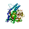
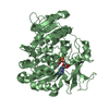
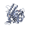
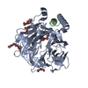
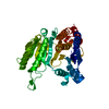
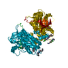
Entry Database : PDB / ID : 4g5jTitle Crystal structure of EGFR kinase in complex with BIBW2992 Epidermal growth factor receptor Keywords / / Function / homology Function Domain/homology Component
/ / / / / / / / / / / / / / / / / / / / / / / / / / / / / / / / / / / / / / / / / / / / / / / / / / / / / / / / / / / / / / / / / / / / / / / / / / / / / / / / / / / / / / / / / / / / / / / / / / / / / / / / / / / / / / / / / / / / / / / / / / / / / / / / / / / / / / / / / / / / / / / / / / / / / Biological species Homo sapiens (human)Method / / Resolution : 2.8 Å Authors Solca, F. / Dahl, G. / Zoephel, A. / Bader, G. / Sanderson, M. / Klein, C. / Kraemer, O. / Himmelsbach, F. / Haaksma, E. / Adolf, G.R. Journal : J.Pharmacol.Exp.Ther. / Year : 2012Title : Target Binding Properties and Cellular Activity of Afatinib (BIBW 2992), an Irreversible ErbB Family Blocker.Authors : Solca, F. / Dahl, G. / Zoephel, A. / Bader, G. / Sanderson, M. / Klein, C. / Kraemer, O. / Himmelsbach, F. / Haaksma, E. / Adolf, G.R. History Deposition Jul 18, 2012 Deposition site / Processing site Revision 1.0 Aug 29, 2012 Provider / Type Revision 1.1 Oct 31, 2012 Group Revision 1.2 Nov 6, 2024 Group Data collection / Database references ... Data collection / Database references / Derived calculations / Structure summary Category chem_comp_atom / chem_comp_bond ... chem_comp_atom / chem_comp_bond / database_2 / pdbx_entry_details / pdbx_modification_feature / struct_conn / struct_site Item _database_2.pdbx_DOI / _database_2.pdbx_database_accession ... _database_2.pdbx_DOI / _database_2.pdbx_database_accession / _struct_conn.pdbx_leaving_atom_flag / _struct_site.pdbx_auth_asym_id / _struct_site.pdbx_auth_comp_id / _struct_site.pdbx_auth_seq_id
Show all Show less
 Movie
Movie Controller


 Open data
 Basic information
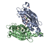
 Components
 Keywords
 Function and homology information
 Homo sapiens (human)
 X-RAY DIFFRACTION /
  Authors
 Citation
 Structure visualization
 Downloads & links
 Other downloads
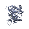
 PDBj
PDBj


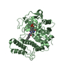
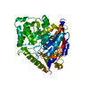
 Assembly
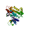
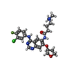
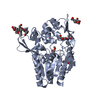
 Mass: 487.954 Da / Num. of mol.: 1 / Source method: obtained synthetically / Formula: C24H27ClFN5O3 / Comment: medication, inhibitor*YM
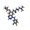
 Mass: 485.938 Da / Num. of mol.: 2 / Source method: obtained synthetically / Formula: C24H25ClFN5O3 / Comment: medication, inhibitor*YM
 Mass: 18.015 Da / Num. of mol.: 26 / Source method: isolated from a natural source / Formula: H2O
 Sample preparation
 / Beamline: X06SA / Wavelength: 0.9999 / Wavelength: 1 Å
 Processing