 Movie
Movie Controller

[English] 日本語
 Yorodumi
- PDB-2xqa: Pentameric ligand gated ion channel GLIC in complex with tetrabut... -
+ Open data

- Basic information
| Entry | Database: PDB / ID: 2xqa | ||||||
|---|---|---|---|---|---|---|---|
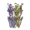
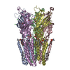
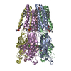
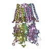
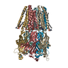
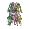
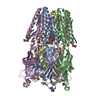
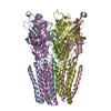
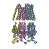
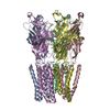
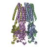
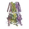
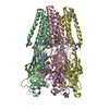
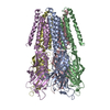
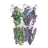
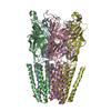
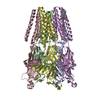
| Title | Pentameric ligand gated ion channel GLIC in complex with tetrabutylantimony (TBSb) | ||||||
 Components Components | GLR4197 PROTEIN | ||||||
 Keywords Keywords | MEMBRANE PROTEIN / OPEN CHANNEL BLOCK | ||||||
| Function / homology |  Function and homology information Function and homology informationsodium channel activity / potassium channel activity / extracellular ligand-gated monoatomic ion channel activity / transmembrane signaling receptor activity / identical protein binding / plasma membrane Similarity search - Function | ||||||
| Biological species |  GLOEOBACTER VIOLACEUS PCC 7421 (bacteria) GLOEOBACTER VIOLACEUS PCC 7421 (bacteria) | ||||||
| Method |  X-RAY DIFFRACTION / SYNCHROTRON / OTHER / Resolution: 3.7 Å X-RAY DIFFRACTION / SYNCHROTRON / OTHER / Resolution: 3.7 Å | ||||||
 Authors Authors | Hilf, R.J.C. / Bertozzi, C. / Zimmermann, I. / Reiter, A. / Trauner, D. / Dutzler, R. | ||||||
 Citation Citation | Journal: Nat.Struct.Mol.Biol. / Year: 2010 Title: Structural Basis of Open Channel Block in a Prokaryotic Pentameric Ligand-Gated Ion Channel Authors: Hilf, R.J.C. / Bertozzi, C. / Zimmermann, I. / Reiter, A. / Trauner, D. / Dutzler, R. #1: Journal: Nature / Year: 2009Title: Structure of a Potentially Open State of a Proton-Activated Pentameric Ligand-Gated Ion Channel. Authors: Hilf, R.J.C. / Dutzler, R. | ||||||
| History |
|
- Structure visualization
| Structure viewer | Molecule: MolmilJmol/JSmol |
|---|
- Downloads & links
-Download
| PDBx/mmCIF format | 2xqa.cif.gz | 624.3 KB | Display | PDBx/mmCIF format |
|---|---|---|---|---|
| PDB format | pdb2xqa.ent.gz | 523.2 KB | Display | PDB format |
| PDBx/mmJSON format | 2xqa.json.gz | Tree view | PDBx/mmJSON format | |
| Others |  Other downloads Other downloads |
-Validation report
| Arichive directory | https://data.pdbj.org/pub/pdb/validation_reports/xq/2xqaftp://data.pdbj.org/pub/pdb/validation_reports/xq/2xqa | HTTPS FTP |
|---|
-Related structure data
| Related structure data |  2xq3C  2xq4C  2xq5C  2xq6C  2xq7C  2xq8C  2xq9C C: citing same article ( |
|---|---|
| Similar structure data |


-Links
 PDBj
PDBj

- Assembly
| Deposited unit | 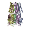
| ||||||||||||||||||||||||||||||||||||
|---|---|---|---|---|---|---|---|---|---|---|---|---|---|---|---|---|---|---|---|---|---|---|---|---|---|---|---|---|---|---|---|---|---|---|---|---|---|
| 1 |
| ||||||||||||||||||||||||||||||||||||
| Unit cell |
| ||||||||||||||||||||||||||||||||||||
| Noncrystallographic symmetry (NCS) | NCS domain:
NCS domain segments:
|
-Components
| #1: Protein | Mass: 36277.723 Da / Num. of mol.: 5 / Fragment: RESIDUES 43-359 Source method: isolated from a genetically manipulated source Details: SB ION Source: (gene. exp.) GLOEOBACTER VIOLACEUS PCC 7421 (bacteria)Plasmid: PET26 / Production host: #2: Chemical | ChemComp-SB / |   Mass: 121.760 Da / Num. of mol.: 1 / Source method: obtained synthetically / Formula: Sb Mass: 121.760 Da / Num. of mol.: 1 / Source method: obtained synthetically / Formula: SbSequence details | RESIDUES 50-359 | |
|---|
-Experimental details
-Experiment
| Experiment | Method: X-RAY DIFFRACTION / Number of used crystals: 1 |
|---|
- Sample preparation
| Crystal | Density Matthews: 5.31 Å3/Da / Density % sol: 76.67 % / Description: NONE |
|---|---|
| Crystal grow | Temperature: 277.15 K / Method: vapor diffusion, hanging drop / pH: 4 Details: 9% PEG4000, 50MM CH3COONA, 200MM (NH4)2SO4, PH4.0, VAPOR DIFFUSION, HANGING DROP, TEMPERATURE 277.15K, VAPOR DIFFUSION, HANGING DROP |
-Data collection
| Diffraction | Mean temperature: 277.15 K |
|---|---|
| Diffraction source | Source: SYNCHROTRON / Site: SLS  / Beamline: X06SA / Wavelength: 1.8 / Beamline: X06SA / Wavelength: 1.8 |
| Detector | Type: DECTRIS PILATUS 6M / Detector: PIXEL / Date: Oct 15, 2008 |
| Radiation | Protocol: SINGLE WAVELENGTH / Monochromatic (M) / Laue (L): M / Scattering type: x-ray |
| Radiation wavelength | Wavelength: 1.8 Å / Relative weight: 1 |
| Reflection | Resolution: 3.6→40.2 Å / Num. obs: 43105 / % possible obs: 99.4 % / Observed criterion σ(I): 2.2 / Redundancy: 5.6 % / Biso Wilson estimate: 92.5 Å2 / Rmerge(I) obs: 0.09 |
- Processing
| Software |
| |||||||||||||||||||||||||||||||||||||||||||||||||||||||||||||||||||||||||||||||||||||||||||||||||||||||||
|---|---|---|---|---|---|---|---|---|---|---|---|---|---|---|---|---|---|---|---|---|---|---|---|---|---|---|---|---|---|---|---|---|---|---|---|---|---|---|---|---|---|---|---|---|---|---|---|---|---|---|---|---|---|---|---|---|---|---|---|---|---|---|---|---|---|---|---|---|---|---|---|---|---|---|---|---|---|---|---|---|---|---|---|---|---|---|---|---|---|---|---|---|---|---|---|---|---|---|---|---|---|---|---|---|---|---|
| Refinement | Method to determine structure: OTHER Starting model: NONE Resolution: 3.7→40.2 Å / SU ML: 0.59 / σ(F): 1.9 / Phase error: 31.41 / Stereochemistry target values: ML
| |||||||||||||||||||||||||||||||||||||||||||||||||||||||||||||||||||||||||||||||||||||||||||||||||||||||||
| Solvent computation | Shrinkage radii: 0.9 Å / VDW probe radii: 1.11 Å / Solvent model: FLAT BULK SOLVENT MODEL / Bsol: 50.467 Å2 / ksol: 0.276 e/Å3 | |||||||||||||||||||||||||||||||||||||||||||||||||||||||||||||||||||||||||||||||||||||||||||||||||||||||||
| Displacement parameters |
| |||||||||||||||||||||||||||||||||||||||||||||||||||||||||||||||||||||||||||||||||||||||||||||||||||||||||
| Refinement step | Cycle: LAST / Resolution: 3.7→40.2 Å
| |||||||||||||||||||||||||||||||||||||||||||||||||||||||||||||||||||||||||||||||||||||||||||||||||||||||||
| Refine LS restraints |
| |||||||||||||||||||||||||||||||||||||||||||||||||||||||||||||||||||||||||||||||||||||||||||||||||||||||||
| Refine LS restraints NCS |
| |||||||||||||||||||||||||||||||||||||||||||||||||||||||||||||||||||||||||||||||||||||||||||||||||||||||||
| LS refinement shell |
| |||||||||||||||||||||||||||||||||||||||||||||||||||||||||||||||||||||||||||||||||||||||||||||||||||||||||
| Refinement TLS params. | Method: refined / Origin x: 24.9164 Å / Origin y: 3.1883 Å / Origin z: 52.6059 Å
| |||||||||||||||||||||||||||||||||||||||||||||||||||||||||||||||||||||||||||||||||||||||||||||||||||||||||
| Refinement TLS group | Selection details: ALL |
