 Movie
Movie Controller

[English] 日本語
 Yorodumi
- PDB-3uu4: The GLIC pentameric Ligand-Gated Ion Channel Loop2-21' mutant red... -
+ Open data

- Basic information
| Entry | Database: PDB / ID: 3uu4 | ||||||
|---|---|---|---|---|---|---|---|
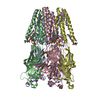
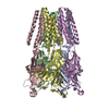
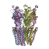
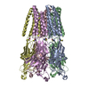
| Title | The GLIC pentameric Ligand-Gated Ion Channel Loop2-21' mutant reduced in the crystal in a locally-closed conformation (LC1 subtype) | ||||||
 Components Components | Glr4197 protein | ||||||
 Keywords Keywords | MEMBRANE PROTEIN / TRANSPORT PROTEIN / Cys-loop receptor family | ||||||
| Function / homology |  Function and homology information Function and homology informationsodium channel activity / potassium channel activity / extracellular ligand-gated monoatomic ion channel activity / transmembrane signaling receptor activity / identical protein binding / plasma membrane Similarity search - Function | ||||||
| Biological species |  Gloeobacter violaceus (bacteria) Gloeobacter violaceus (bacteria) | ||||||
| Method |  X-RAY DIFFRACTION / SYNCHROTRON / MOLECULAR REPLACEMENT / Resolution: 3.05 Å X-RAY DIFFRACTION / SYNCHROTRON / MOLECULAR REPLACEMENT / Resolution: 3.05 Å | ||||||
 Authors Authors | Sauguet, L. / Nury, H. / Corringer, P.J. / Delarue, M. | ||||||
 Citation Citation | Journal: Nat.Struct.Mol.Biol. / Year: 2012 Title: A locally closed conformation of a bacterial pentameric proton-gated ion channel. Authors: Prevost, M.S. / Sauguet, L. / Nury, H. / Van Renterghem, C. / Huon, C. / Poitevin, F. / Baaden, M. / Delarue, M. / Corringer, P.J. | ||||||
| History |
|
- Structure visualization
| Structure viewer | Molecule: MolmilJmol/JSmol |
|---|
- Downloads & links
-Download
| PDBx/mmCIF format | 3uu4.cif.gz | 313.8 KB | Display | PDBx/mmCIF format |
|---|---|---|---|---|
| PDB format | pdb3uu4.ent.gz | 257.8 KB | Display | PDB format |
| PDBx/mmJSON format | 3uu4.json.gz | Tree view | PDBx/mmJSON format | |
| Others |  Other downloads Other downloads |
-Validation report
| Arichive directory | https://data.pdbj.org/pub/pdb/validation_reports/uu/3uu4ftp://data.pdbj.org/pub/pdb/validation_reports/uu/3uu4 | HTTPS FTP |
|---|
-Related structure data
| Related structure data |  3tlsC  3tltC  3tluC  3tlvC  3tlwC  3uu3C  3uu5C  3uu6C  3uu8C  3uubC  3eamS C: citing same article ( S: Starting model for refinement |
|---|---|
| Similar structure data |
-Links
 PDBj
PDBj

- Assembly
| Deposited unit | 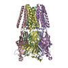
| ||||||||
|---|---|---|---|---|---|---|---|---|---|
| 1 |
| ||||||||
| Unit cell |
| ||||||||
| Components on special symmetry positions |
|
-Components
| #1: Protein | Mass: 36524.977 Da / Num. of mol.: 5 / Fragment: UNP residues 44-359 / Mutation: C27S,K33C,N245C Source method: isolated from a genetically manipulated source Source: (gene. exp.) Gloeobacter violaceus (bacteria) / Gene: glr4197 / Production host: #2: Sugar | ChemComp-LMT / |   Type: D-saccharide / Mass: 510.615 Da / Num. of mol.: 1 / Source method: obtained synthetically / Formula: C24H46O11 / Comment: detergent*YM Type: D-saccharide / Mass: 510.615 Da / Num. of mol.: 1 / Source method: obtained synthetically / Formula: C24H46O11 / Comment: detergent*YM#3: Water | ChemComp-HOH / |  Mass: 18.015 Da / Num. of mol.: 51 / Source method: isolated from a natural source / Formula: H2O Mass: 18.015 Da / Num. of mol.: 51 / Source method: isolated from a natural source / Formula: H2O |
|---|
-Experimental details
-Experiment
| Experiment | Method: X-RAY DIFFRACTION / Number of used crystals: 1 |
|---|
- Sample preparation
| Crystal | Density Matthews: 5.2 Å3/Da / Density % sol: 76.34 % |
|---|---|
| Crystal grow | Temperature: 293 K / Method: vapor diffusion, hanging drop / pH: 4 Details: 15% PEG4000, 0.4 M sodium thiocyanate, 0.1 M sodium acetate, pH 4.0, VAPOR DIFFUSION, HANGING DROP, temperature 293K |
-Data collection
| Diffraction | Mean temperature: 100 K | ||||||||||||||||||
|---|---|---|---|---|---|---|---|---|---|---|---|---|---|---|---|---|---|---|---|
| Diffraction source | Source: SYNCHROTRON / Site: SOLEIL  / Beamline: PROXIMA 1 / Wavelength: 0.98011 Å / Beamline: PROXIMA 1 / Wavelength: 0.98011 Å | ||||||||||||||||||
| Detector | Type: ADSC QUANTUM 315r / Detector: CCD / Date: Feb 2, 2011 | ||||||||||||||||||
| Radiation | Monochromator: Si(111) / Protocol: SINGLE WAVELENGTH / Monochromatic (M) / Laue (L): M / Scattering type: x-ray | ||||||||||||||||||
| Radiation wavelength | Wavelength: 0.98011 Å / Relative weight: 1 | ||||||||||||||||||
| Reflection | Resolution: 3.05→40.9 Å / Num. all: 71200 / Num. obs: 70985 / % possible obs: 99.7 % / Redundancy: 3.7 % / Biso Wilson estimate: 90.4 Å2 / Rmerge(I) obs: 0.07 / Rsym value: 0.043 / Net I/σ(I): 10.8 | ||||||||||||||||||
| Reflection shell | Diffraction-ID: 1 / Redundancy: 3.7 %
|
- Processing
| Software |
| ||||||||||||||||||||||||||||||||||||||||||||||||||||||||||||||||||||||||||||||||||||||||||||||||||||||||||||
|---|---|---|---|---|---|---|---|---|---|---|---|---|---|---|---|---|---|---|---|---|---|---|---|---|---|---|---|---|---|---|---|---|---|---|---|---|---|---|---|---|---|---|---|---|---|---|---|---|---|---|---|---|---|---|---|---|---|---|---|---|---|---|---|---|---|---|---|---|---|---|---|---|---|---|---|---|---|---|---|---|---|---|---|---|---|---|---|---|---|---|---|---|---|---|---|---|---|---|---|---|---|---|---|---|---|---|---|---|---|
| Refinement | Method to determine structure: MOLECULAR REPLACEMENT Starting model: PDB ENTRY 3EAM Resolution: 3.05→27.28 Å / Cor.coef. Fo:Fc: 0.8574 / Cor.coef. Fo:Fc free: 0.8392 / SU R Cruickshank DPI: 0.531 / Cross valid method: THROUGHOUT / σ(F): 0 / SU R Blow DPI: 0.473 / SU Rfree Blow DPI: 0.296 / SU Rfree Cruickshank DPI: 0.311
| ||||||||||||||||||||||||||||||||||||||||||||||||||||||||||||||||||||||||||||||||||||||||||||||||||||||||||||
| Displacement parameters | Biso mean: 104.69 Å2
| ||||||||||||||||||||||||||||||||||||||||||||||||||||||||||||||||||||||||||||||||||||||||||||||||||||||||||||
| Refine analyze | Luzzati coordinate error obs: 0.71 Å | ||||||||||||||||||||||||||||||||||||||||||||||||||||||||||||||||||||||||||||||||||||||||||||||||||||||||||||
| Refinement step | Cycle: LAST / Resolution: 3.05→27.28 Å
| ||||||||||||||||||||||||||||||||||||||||||||||||||||||||||||||||||||||||||||||||||||||||||||||||||||||||||||
| Refine LS restraints |
| ||||||||||||||||||||||||||||||||||||||||||||||||||||||||||||||||||||||||||||||||||||||||||||||||||||||||||||
| LS refinement shell | Resolution: 3.05→3.13 Å / Total num. of bins used: 20
|
