[English] 日本語
Yorodumi
- PDB-2xhz: Probing the active site of the sugar isomerase domain from E. col... -

+
Open data


ID or keywords:

Loading...

-
Basic information

Entry
Database: PDB / ID: 2xhz
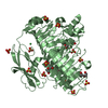
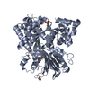
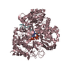
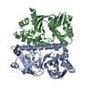
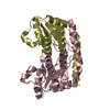
TitleProbing the active site of the sugar isomerase domain from E. coli arabinose-5-phosphate isomerase via X-ray crystallography
ComponentsARABINOSE 5-PHOSPHATE ISOMERASE
KeywordsISOMERASE / LIPOPOLYSACCHARIDE BIOGENESIS
Function / homology
Function and homology information


arabinose-5-phosphate isomerase / arabinose-5-phosphate isomerase activity / keto-3-deoxy-D-manno-octulosonic acid biosynthetic process / carbohydrate derivative binding / metal ion binding
Similarity search - Function
Phosphosugar isomerase, KdsD/KpsF-type / KpsF-like, SIS domain / SIS domain / SIS domain / SIS domain profile. / Glucose-6-phosphate isomerase like protein; domain 1 / CBS domain / CBS domain / CBS domain profile. / Rossmann fold ...Phosphosugar isomerase, KdsD/KpsF-type / KpsF-like, SIS domain / SIS domain / SIS domain / SIS domain profile. / Glucose-6-phosphate isomerase like protein; domain 1 / CBS domain / CBS domain / CBS domain profile. / Rossmann fold / 3-Layer(aba) Sandwich / Alpha Beta
Similarity search - Domain/homology
Arabinose 5-phosphate isomerase KdsD
Similarity search - Component
Biological speciesESCHERICHIA COLI (E. coli)
MethodX-RAY DIFFRACTION / SYNCHROTRON / MOLECULAR REPLACEMENT / Resolution: 2.6 Å
AuthorsGourlay, L.J. / Sommaruga, S. / Nardini, M. / Sperandeo, P. / Deho, G. / Polissi, A. / Bolognesi, M.
CitationJournal: Protein Sci. / Year: 2010
Title: Probing the Active Site of the Sugar Isomerase Domain from E. Coli Arabinose-5-Phosphate Isomerase Via X-Ray Crystallography.
Authors: Gourlay, L.J. / Sommaruga, S. / Nardini, M. / Sperandeo, P. / Deho, G. / Polissi, A. / Bolognesi, M.
History
DepositionJun 24, 2010Deposition site: PDBE / Processing site: PDBE
Revision 1.0Jan 26, 2011Provider: repository / Type: Initial release
Revision 1.1May 8, 2011Group: Version format compliance
Revision 1.2Jul 13, 2011Group: Version format compliance
Revision 1.3Dec 20, 2023Group: Data collection / Database references ...Data collection / Database references / Other / Refinement description
Category: chem_comp_atom / chem_comp_bond ...chem_comp_atom / chem_comp_bond / database_2 / pdbx_database_status / pdbx_initial_refinement_model
Item: _database_2.pdbx_DOI / _database_2.pdbx_database_accession / _pdbx_database_status.status_code_sf

-
Structure visualization

Structure viewerMolecule:
MolmilJmol/JSmol

Downloads & links

-
Assembly

Deposited unit
A: ARABINOSE 5-PHOSPHATE ISOMERASE
B: ARABINOSE 5-PHOSPHATE ISOMERASE
C: ARABINOSE 5-PHOSPHATE ISOMERASE
D: ARABINOSE 5-PHOSPHATE ISOMERASE


Theoretical massNumber of molelcules
Total (without water)77,3184
Polymers77,3184
Non-polymers00
Water1629
1


  • Idetical with deposited unit
  • defined by author&software
TypeNameSymmetry operationNumber
identity operation1_555x,y,z1
Buried area11760 Å2
ΔGint-98.4 kcal/mol
Surface area24210 Å2
MethodPISA
2
A: ARABINOSE 5-PHOSPHATE ISOMERASE
B: ARABINOSE 5-PHOSPHATE ISOMERASE


Theoretical massNumber of molelcules
Total (without water)38,6592
Polymers38,6592
Non-polymers00
Water362
TypeNameSymmetry operationNumber
identity operation1_555x,y,z1
Buried area4230 Å2
ΔGint-38.9 kcal/mol
Surface area13810 Å2
MethodPISA
3
C: ARABINOSE 5-PHOSPHATE ISOMERASE
D: ARABINOSE 5-PHOSPHATE ISOMERASE


Theoretical massNumber of molelcules
Total (without water)38,6592
Polymers38,6592
Non-polymers00
Water362
TypeNameSymmetry operationNumber
identity operation1_555x,y,z1
Buried area4200 Å2
ΔGint-36.8 kcal/mol
Surface area13730 Å2
MethodPISA
Unit cell
Length a, b, c (Å)55.860, 67.430, 82.170
Angle α, β, γ (deg.)90.00, 106.94, 90.00
Int Tables number4
Space group name H-MP1211
Noncrystallographic symmetry (NCS)NCS domain:
IDEns-IDDetails
11A
21B
31C
41D

NCS domain segments:
Dom-IDComponent-IDEns-IDRefine codeAuth asym-IDAuth seq-ID
1114A10 - 183
2114B10 - 183
3114C10 - 183
4114D10 - 183

-
Components

#1: Protein
ARABINOSE 5-PHOSPHATE ISOMERASE / KDSD / YRBH


Mass: 19329.508 Da / Num. of mol.: 4 / Fragment: SUGAR ISOMERASE DOMAIN, RESIDUES 1-183 / Mutation: YES
Source method: isolated from a genetically manipulated source
Source: (gene. exp.) ESCHERICHIA COLI (E. coli) / Production host: ESCHERICHIA COLI (E. coli) / Strain (production host): BL21
References: UniProt: P45395, arabinose-5-phosphate isomerase
#2: Water ChemComp-HOH / water


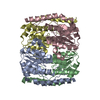
Mass: 18.015 Da / Num. of mol.: 9 / Source method: isolated from a natural source / Formula: H2O

-
Experimental details

-
Experiment

ExperimentMethod: X-RAY DIFFRACTION / Number of used crystals: 1

-
Sample preparation

CrystalDensity Matthews: 1.8 Å3/Da / Density % sol: 32 % / Description: NONE
Crystal growpH: 7.5
Details: 20% PEG-8000 100MM HEPES PH 7.5 (WIZARD I SCREEN,[EMERALD BIOSYSTEMS] CONDITION 21).

-
Data collection

DiffractionMean temperature: 100 K
Diffraction sourceSource: SYNCHROTRON / Site: ESRF / Beamline: ID23-1 / Wavelength: 1
DetectorType: ADSC QUANTUM 315r / Detector: CCD / Date: Jun 25, 2009
RadiationProtocol: SINGLE WAVELENGTH / Monochromatic (M) / Laue (L): M / Scattering type: x-ray
Radiation wavelengthWavelength: 1 Å / Relative weight: 1
ReflectionResolution: 2.6→40 Å / Num. obs: 17026 / % possible obs: 94.3 % / Observed criterion σ(I): 2 / Redundancy: 2.7 % / Biso Wilson estimate: 42.7 Å2 / Rmerge(I) obs: 0.12 / Net I/σ(I): 5.9
Reflection shellResolution: 2.6→2.74 Å / Redundancy: 2.7 % / Rmerge(I) obs: 0.36 / Mean I/σ(I) obs: 2.4 / % possible all: 95.5

-
Processing

Software
NameVersionClassification
REFMAC5.5.0066refinement
iMOSFLMdata reduction
SCALAdata scaling
MOLREPphasing
RefinementMethod to determine structure: MOLECULAR REPLACEMENT
Starting model: PDB ENTRY 3ETN
Resolution: 2.6→40 Å / Cor.coef. Fo:Fc: 0.895 / Cor.coef. Fo:Fc free: 0.858 / SU B: 20.197 / SU ML: 0.409 / Cross valid method: THROUGHOUT / ESU R Free: 0.463 / Stereochemistry target values: MAXIMUM LIKELIHOOD / Details: HYDROGENS HAVE BEEN ADDED IN THE RIDING POSITIONS.
RfactorNum. reflection% reflectionSelection details
Rfree0.30503 863 5.1 %RANDOM
Rwork0.26075 ---
obs0.26302 16148 93.68 %-
Solvent computationIon probe radii: 0.8 Å / Shrinkage radii: 0.8 Å / VDW probe radii: 1.4 Å / Solvent model: MASK
Displacement parametersBiso mean: 36.384 Å2
Baniso -1Baniso -2Baniso -3
1-5.16 Å20 Å25.24 Å2
2---2.76 Å20 Å2
3---0.66 Å2
Refinement stepCycle: LAST / Resolution: 2.6→40 Å
ProteinNucleic acidLigandSolventTotal
Num. atoms5086 0 0 9 5095
Refine LS restraints
Refine-IDTypeDev idealDev ideal targetNumber
X-RAY DIFFRACTIONr_bond_refined_d0.0020.0225172
X-RAY DIFFRACTIONr_bond_other_d
X-RAY DIFFRACTIONr_angle_refined_deg0.431.977003
X-RAY DIFFRACTIONr_angle_other_deg
X-RAY DIFFRACTIONr_dihedral_angle_1_deg2.8135689
X-RAY DIFFRACTIONr_dihedral_angle_2_deg24.86124.138174
X-RAY DIFFRACTIONr_dihedral_angle_3_deg11.6115882
X-RAY DIFFRACTIONr_dihedral_angle_4_deg15.5071524
X-RAY DIFFRACTIONr_chiral_restr0.0310.2838
X-RAY DIFFRACTIONr_gen_planes_refined
X-RAY DIFFRACTIONr_gen_planes_other
X-RAY DIFFRACTIONr_nbd_refined
X-RAY DIFFRACTIONr_nbd_other
X-RAY DIFFRACTIONr_nbtor_refined
X-RAY DIFFRACTIONr_nbtor_other
X-RAY DIFFRACTIONr_xyhbond_nbd_refined
X-RAY DIFFRACTIONr_xyhbond_nbd_other
X-RAY DIFFRACTIONr_metal_ion_refined
X-RAY DIFFRACTIONr_metal_ion_other
X-RAY DIFFRACTIONr_symmetry_vdw_refined
X-RAY DIFFRACTIONr_symmetry_vdw_other
X-RAY DIFFRACTIONr_symmetry_hbond_refined
X-RAY DIFFRACTIONr_symmetry_hbond_other
X-RAY DIFFRACTIONr_symmetry_metal_ion_refined
X-RAY DIFFRACTIONr_symmetry_metal_ion_other
X-RAY DIFFRACTIONr_mcbond_it0.4431.53434
X-RAY DIFFRACTIONr_mcbond_other
X-RAY DIFFRACTIONr_mcangle_it0.8225499
X-RAY DIFFRACTIONr_mcangle_other
X-RAY DIFFRACTIONr_scbond_it0.71131738
X-RAY DIFFRACTIONr_scbond_other
X-RAY DIFFRACTIONr_scangle_it1.3624.51504
X-RAY DIFFRACTIONr_scangle_other
X-RAY DIFFRACTIONr_long_range_B_refined
X-RAY DIFFRACTIONr_long_range_B_other
X-RAY DIFFRACTIONr_rigid_bond_restr
X-RAY DIFFRACTIONr_sphericity_free
X-RAY DIFFRACTIONr_sphericity_bonded
Refine LS restraints NCS

Ens-ID: 1 / Number: 1255 / Refine-ID: X-RAY DIFFRACTION

Dom-IDAuth asym-IDTypeRms dev position (Å)Weight position
1Amedium positional0.420.5
2Bmedium positional0.420.5
3Cmedium positional0.390.5
4Dmedium positional0.370.5
1Amedium thermal0.052
2Bmedium thermal0.052
3Cmedium thermal0.052
4Dmedium thermal0.052
LS refinement shellResolution: 2.6→2.667 Å / Total num. of bins used: 20
RfactorNum. reflection% reflection
Rfree0.364 57 -
Rwork0.362 1235 -
obs--95.56 %

+
About Yorodumi

-
News

-
Feb 9, 2022. New format data for meta-information of EMDB entries

New format data for meta-information of EMDB entries

  • Version 3 of the EMDB header file is now the official format.
  • The previous official version 1.9 will be removed from the archive.

Related info.:EMDB header

External links:wwPDB to switch to version 3 of the EMDB data model

-
Aug 12, 2020. Covid-19 info

Covid-19 info

URL: https://pdbjlvh1.pdbj.org/emnavi/covid19.php

New page: Covid-19 featured information page in EM Navigator.

Related info.:Covid-19 info / Mar 5, 2020. Novel coronavirus structure data

+
Mar 5, 2020. Novel coronavirus structure data

Novel coronavirus structure data

Related info.:Yorodumi Speices / Aug 12, 2020. Covid-19 info

External links:COVID-19 featured content - PDBj / Molecule of the Month (242):Coronavirus Proteases

+
Jan 31, 2019. EMDB accession codes are about to change! (news from PDBe EMDB page)

EMDB accession codes are about to change! (news from PDBe EMDB page)

  • The allocation of 4 digits for EMDB accession codes will soon come to an end. Whilst these codes will remain in use, new EMDB accession codes will include an additional digit and will expand incrementally as the available range of codes is exhausted. The current 4-digit format prefixed with “EMD-” (i.e. EMD-XXXX) will advance to a 5-digit format (i.e. EMD-XXXXX), and so on. It is currently estimated that the 4-digit codes will be depleted around Spring 2019, at which point the 5-digit format will come into force.
  • The EM Navigator/Yorodumi systems omit the EMD- prefix.

Related info.:Q: What is EMD? / ID/Accession-code notation in Yorodumi/EM Navigator

External links:EMDB Accession Codes are Changing Soon! / Contact to PDBj

+
Jul 12, 2017. Major update of PDB

Major update of PDB

  • wwPDB released updated PDB data conforming to the new PDBx/mmCIF dictionary.
  • This is a major update changing the version number from 4 to 5, and with Remediation, in which all the entries are updated.
  • In this update, many items about electron microscopy experimental information are reorganized (e.g. em_software).
  • Now, EM Navigator and Yorodumi are based on the updated data.

External links:wwPDB Remediation / Enriched Model Files Conforming to OneDep Data Standards Now Available in the PDB FTP Archive

-
Yorodumi

Thousand views of thousand structures

  • Yorodumi is a browser for structure data from EMDB, PDB, SASBDB, etc.
  • This page is also the successor to EM Navigator detail page, and also detail information page/front-end page for Omokage search.
  • The word "yorodu" (or yorozu) is an old Japanese word meaning "ten thousand". "mi" (miru) is to see.

Related info.:EMDB / PDB / SASBDB / Comparison of 3 databanks / Yorodumi Search / Aug 31, 2016. New EM Navigator & Yorodumi / Yorodumi Papers / Jmol/JSmol / Function and homology information / Changes in new EM Navigator and Yorodumi

Read more