 Movie
Movie Controller

[English] 日本語
 Yorodumi
- PDB-2xf1: Crystal structure of Plasmodium falciparum actin depolymerization... -
+ Open data

- Basic information
| Entry | Database: PDB / ID: 2xf1 | ||||||
|---|---|---|---|---|---|---|---|
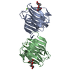
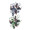
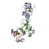
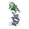
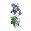
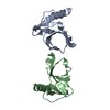
| Title | Crystal structure of Plasmodium falciparum actin depolymerization factor 1 | ||||||
 Components Components | COFILIN ACTIN-DEPOLYMERIZING FACTOR HOMOLOG 1 | ||||||
 Keywords Keywords | ACTIN-BINDING PROTEIN / CYTOSKELETON | ||||||
| Function / homology |  Function and homology information Function and homology informationcytoplasmic actin-based contraction involved in cell motility / actin filament severing / actin filament depolymerization / actin monomer binding / actin filament binding / actin cytoskeleton / actin cytoskeleton organization / cytoplasm / cytosol Similarity search - Function | ||||||
| Biological species |  | ||||||
| Method |  X-RAY DIFFRACTION / SYNCHROTRON / MOLECULAR REPLACEMENT / Resolution: 1.96 Å X-RAY DIFFRACTION / SYNCHROTRON / MOLECULAR REPLACEMENT / Resolution: 1.96 Å | ||||||
 Authors Authors | Singh, B.K. / Sattler, J.M. / Huttu, J. / Chatterjee, M. / Schueler, H. / Kursula, I. | ||||||
 Citation Citation | Journal: J.Biol.Chem. / Year: 2011 Title: Crystal Structures Explain Functional Differences in the Two Actin Depolymerization Factors of the Malaria Parasite. Authors: Singh, B.K. / Sattler, J.M. / Chatterjee, M. / Huttu, J. / Schuler, H. / Kursula, I. | ||||||
| History |
|
- Structure visualization
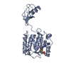
| Structure viewer | Molecule: MolmilJmol/JSmol |
|---|
- Downloads & links
-Download
| PDBx/mmCIF format | 2xf1.cif.gz | 67.6 KB | Display | PDBx/mmCIF format |
|---|---|---|---|---|
| PDB format | pdb2xf1.ent.gz | 50.3 KB | Display | PDB format |
| PDBx/mmJSON format | 2xf1.json.gz | Tree view | PDBx/mmJSON format | |
| Others |  Other downloads Other downloads |
-Validation report
| Summary document | 2xf1_validation.pdf.gz | 442.6 KB | Display | wwPDB validaton report |
|---|---|---|---|---|
| Full document | 2xf1_full_validation.pdf.gz | 444.3 KB | Display | |
| Data in XML | 2xf1_validation.xml.gz | 9.8 KB | Display | |
| Data in CIF | 2xf1_validation.cif.gz | 13.7 KB | Display | |
| Arichive directory | https://data.pdbj.org/pub/pdb/validation_reports/xf/2xf1ftp://data.pdbj.org/pub/pdb/validation_reports/xf/2xf1 | HTTPS FTP |
-Related structure data
| Related structure data |  2xfaC  1ahqS  1f7sS  2i2qS S: Starting model for refinement C: citing same article ( |
|---|---|
| Similar structure data |

-Links
 PDBj
PDBj
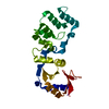
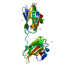
- Assembly
| Deposited unit | 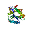
| ||||||||
|---|---|---|---|---|---|---|---|---|---|
| 1 | 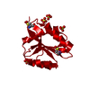
| ||||||||
| Unit cell |
| ||||||||
| Components on special symmetry positions |
|
-Components
| #1: Protein | Mass: 14184.280 Da / Num. of mol.: 1 Source method: isolated from a genetically manipulated source Details: CYS24 AND CYS34 PARTIALLY OXIDIZED Source: (gene. exp.) Plasmid: PGEX-6P-1 / Production host:  | ||||
|---|---|---|---|---|---|
| #2: Chemical | ChemComp-SO4 /   Mass: 96.063 Da / Num. of mol.: 4 / Source method: obtained synthetically / Formula: SO4 Mass: 96.063 Da / Num. of mol.: 4 / Source method: obtained synthetically / Formula: SO4#3: Water | ChemComp-HOH / |  Mass: 18.015 Da / Num. of mol.: 187 / Source method: isolated from a natural source / Formula: H2O Mass: 18.015 Da / Num. of mol.: 187 / Source method: isolated from a natural source / Formula: H2OHas protein modification | Y | |
-Experimental details
-Experiment
| Experiment | Method: X-RAY DIFFRACTION / Number of used crystals: 1 |
|---|
- Sample preparation
| Crystal | Density Matthews: 3.71 Å3/Da / Density % sol: 67 % / Description: NONE |
|---|---|
| Crystal grow | Temperature: 277 K / pH: 7.8 / Details: 2.5 M AMMONUIM SULFATE, 0.1 M HEPES PH 7.8, 277 K |
-Data collection
| Diffraction | Mean temperature: 100 K |
|---|---|
| Diffraction source | Source: SYNCHROTRON / Site: MAX II  / Beamline: I911-2 / Wavelength: 1.038 / Beamline: I911-2 / Wavelength: 1.038 |
| Detector | Type: MARRESEARCH / Detector: CCD / Date: Feb 11, 2010 / Details: MULTILAYER MIRROR |
| Radiation | Monochromator: BENT SI (111) CRYSTAL, HORIZONTALLY FOCUSING / Protocol: SINGLE WAVELENGTH / Monochromatic (M) / Laue (L): M / Scattering type: x-ray |
| Radiation wavelength | Wavelength: 1.038 Å / Relative weight: 1 |
| Reflection | Resolution: 2→20 Å / Num. obs: 14010 / % possible obs: 96.5 % / Observed criterion σ(I): -3 / Redundancy: 10.4 % / Biso Wilson estimate: 24.85 Å2 / Rmerge(I) obs: 0.05 / Net I/σ(I): 38.5 |
| Reflection shell | Resolution: 2→2.05 Å / Redundancy: 7 % / Rmerge(I) obs: 0.28 / Mean I/σ(I) obs: 14.5 / % possible all: 94.5 |
- Processing
| Software |
| ||||||||||||||||||||||||||||||||||||||||||||||||||||||||||||||||||||||||||||||||||||||||||||||||||||||||||||||||||||||||||||||||||||||||||||||||||||||
|---|---|---|---|---|---|---|---|---|---|---|---|---|---|---|---|---|---|---|---|---|---|---|---|---|---|---|---|---|---|---|---|---|---|---|---|---|---|---|---|---|---|---|---|---|---|---|---|---|---|---|---|---|---|---|---|---|---|---|---|---|---|---|---|---|---|---|---|---|---|---|---|---|---|---|---|---|---|---|---|---|---|---|---|---|---|---|---|---|---|---|---|---|---|---|---|---|---|---|---|---|---|---|---|---|---|---|---|---|---|---|---|---|---|---|---|---|---|---|---|---|---|---|---|---|---|---|---|---|---|---|---|---|---|---|---|---|---|---|---|---|---|---|---|---|---|---|---|---|---|---|---|
| Refinement | Method to determine structure: MOLECULAR REPLACEMENT Starting model: PDB ENTRIES 1F7S, 1AHQ AND 2I2Q Resolution: 1.96→27.752 Å / SU ML: 0.29 / σ(F): 1.99 / Phase error: 25.21 / Stereochemistry target values: ML Details: INITIAL STAGES OF REFINEMENT WERE PERFORMED USING REFMAC 5
| ||||||||||||||||||||||||||||||||||||||||||||||||||||||||||||||||||||||||||||||||||||||||||||||||||||||||||||||||||||||||||||||||||||||||||||||||||||||
| Solvent computation | Shrinkage radii: 0.9 Å / VDW probe radii: 1.11 Å / Solvent model: FLAT BULK SOLVENT MODEL / Bsol: 49.275 Å2 / ksol: 0.347 e/Å3 | ||||||||||||||||||||||||||||||||||||||||||||||||||||||||||||||||||||||||||||||||||||||||||||||||||||||||||||||||||||||||||||||||||||||||||||||||||||||
| Displacement parameters |
| ||||||||||||||||||||||||||||||||||||||||||||||||||||||||||||||||||||||||||||||||||||||||||||||||||||||||||||||||||||||||||||||||||||||||||||||||||||||
| Refinement step | Cycle: LAST / Resolution: 1.96→27.752 Å
| ||||||||||||||||||||||||||||||||||||||||||||||||||||||||||||||||||||||||||||||||||||||||||||||||||||||||||||||||||||||||||||||||||||||||||||||||||||||
| Refine LS restraints |
| ||||||||||||||||||||||||||||||||||||||||||||||||||||||||||||||||||||||||||||||||||||||||||||||||||||||||||||||||||||||||||||||||||||||||||||||||||||||
| LS refinement shell |
| ||||||||||||||||||||||||||||||||||||||||||||||||||||||||||||||||||||||||||||||||||||||||||||||||||||||||||||||||||||||||||||||||||||||||||||||||||||||
| Refinement TLS params. | Method: refined / Refine-ID: X-RAY DIFFRACTION
| ||||||||||||||||||||||||||||||||||||||||||||||||||||||||||||||||||||||||||||||||||||||||||||||||||||||||||||||||||||||||||||||||||||||||||||||||||||||
| Refinement TLS group |
|
