 Movie
Movie Controller


+ Open data

- Basic information
| Entry | Database: PDB / ID: 3hip | ||||||
|---|---|---|---|---|---|---|---|
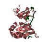
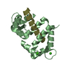
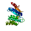
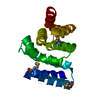
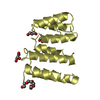
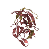
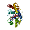
| Title | HIGH-POTENTIAL IRON-SULFUR PROTEIN FROM CHROMATIUM PURPURATUM | ||||||
 Components Components | HIGH-POTENTIAL IRON-SULFUR PROTEIN | ||||||
 Keywords Keywords | ELECTRON TRANSFER / PHOTOSYNTHESIS / METALLOPROTEIN | ||||||
| Function / homology |  Function and homology information Function and homology informationaerobic electron transport chain / 4 iron, 4 sulfur cluster binding / periplasmic space / electron transfer activity / metal ion binding Similarity search - Function | ||||||
| Biological species |  Marichromatium purpuratum (bacteria) Marichromatium purpuratum (bacteria) | ||||||
| Method |  X-RAY DIFFRACTION / MOLECULAR REPLACEMENT / Resolution: 2.8 Å X-RAY DIFFRACTION / MOLECULAR REPLACEMENT / Resolution: 2.8 Å | ||||||
 Authors Authors | Kerfeld, C.A. / Salmeen, A.E. / Yeates, T.O. | ||||||
 Citation Citation | Journal: Biochemistry / Year: 1998 Title: Crystal structure and possible dimerization of the high-potential iron-sulfur protein from Chromatium purpuratum. Authors: Kerfeld, C.A. / Salmeen, A.E. / Yeates, T.O. #1: Journal: Biochemistry / Year: 1996Title: Isolation and Characterization of Soluble Electron Transfer Proteins from Chromatium Purpuratum Authors: Kerfeld, C.A. / Chan, C. / Hirasawa, M. / Kleis-Sanfrancisco, S. / Yeates, T.O. / Knaff, D.B. | ||||||
| History |
|
- Structure visualization
| Structure viewer | Molecule: MolmilJmol/JSmol |
|---|
- Downloads & links
-Download
| PDBx/mmCIF format | 3hip.cif.gz | 54.3 KB | Display | PDBx/mmCIF format |
|---|---|---|---|---|
| PDB format | pdb3hip.ent.gz | 39.7 KB | Display | PDB format |
| PDBx/mmJSON format | 3hip.json.gz | Tree view | PDBx/mmJSON format | |
| Others |  Other downloads Other downloads |
-Validation report
| Arichive directory | https://data.pdbj.org/pub/pdb/validation_reports/hi/3hipftp://data.pdbj.org/pub/pdb/validation_reports/hi/3hip | HTTPS FTP |
|---|
-Related structure data
| Related structure data |  1hipS S: Starting model for refinement |
|---|---|
| Similar structure data |


-Links
 PDBj
PDBj- Assembly
| Deposited unit | 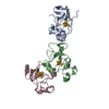
| ||||||||||||
|---|---|---|---|---|---|---|---|---|---|---|---|---|---|
| 1 | 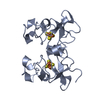
| ||||||||||||
| 2 | 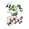
| ||||||||||||
| Unit cell |
| ||||||||||||
| Noncrystallographic symmetry (NCS) | NCS oper:
| ||||||||||||
| Details | MOLECULES B AND C ARE RELATED BY A NONCRYSTALLOGRAPHIC TWO-FOLD SYMMETRY AXIS. |
-Components
| #1: Protein | Mass: 8761.788 Da / Num. of mol.: 3 / Source method: isolated from a natural source / Source: (natural) Marichromatium purpuratum (bacteria) / Cellular location: PERIPLASM / References: UniProt: P59860#2: Chemical |   Mass: 351.640 Da / Num. of mol.: 3 / Source method: obtained synthetically / Formula: Fe4S4 Mass: 351.640 Da / Num. of mol.: 3 / Source method: obtained synthetically / Formula: Fe4S4Sequence details | THE FIRST 25 AMINO ACIDS OF C. PURPURATUM HIPIP WERE DETERMINED BY AUTOMATED EDMAN DEGRADATION (REF. ...THE FIRST 25 AMINO ACIDS OF C. PURPURATUM | |
|---|
-Experimental details
-Experiment
| Experiment | Method: X-RAY DIFFRACTION / Number of used crystals: 2 |
|---|
- Sample preparation
| Crystal | Density Matthews: 1.9 Å3/Da / Density % sol: 34 % | ||||||||||||||||||||
|---|---|---|---|---|---|---|---|---|---|---|---|---|---|---|---|---|---|---|---|---|---|
| Crystal grow | Method: vapor diffusion, hanging drop / pH: 5.4 Details: CRYSTALLIZED BY VAPOR DIFFUSION OVER A RESERVOIR CONTAINING 100 MM CITRATE BUFFER, PH 5.4 AND 3.2 M AMMONIUM SULFATE PROTEIN CONCENTRATION = 8.0 MG/ML, vapor diffusion - hanging drop | ||||||||||||||||||||
| Crystal | *PLUS | ||||||||||||||||||||
| Crystal grow | *PLUS pH: 8 / Method: vapor diffusion, hanging drop | ||||||||||||||||||||
| Components of the solutions | *PLUS
|
-Data collection
| Diffraction | Mean temperature: 298 K |
|---|---|
| Diffraction source | Source: ROTATING ANODE / Type: RIGAKU RUH3R / Wavelength: 1.5418 |
| Detector | Type: RIGAKU / Detector: IMAGE PLATE / Date: Jan 1, 1995 / Details: COLLIMATOR |
| Radiation | Monochromator: GRAPHITE(002) / Monochromatic (M) / Laue (L): M / Scattering type: x-ray |
| Radiation wavelength | Wavelength: 1.5418 Å / Relative weight: 1 |
| Reflection | Highest resolution: 2.7 Å / Num. obs: 6568 / % possible obs: 94.1 % / Observed criterion σ(I): 0 / Redundancy: 5.7 % / Biso Wilson estimate: 24.9 Å2 / Rmerge(I) obs: 0.116 / Net I/σ(I): 6.9 |
| Reflection shell | Resolution: 2.8→2.91 Å / Redundancy: 1.9 % / Rmerge(I) obs: 0.234 / Mean I/σ(I) obs: 3.3 / % possible all: 93 |
| Reflection | *PLUS Lowest resolution: 8 Å / Num. measured all: 13850 |
- Processing
| Software |
| ||||||||||||||||||||||||||||||||||||||||||||||||||||||||||||
|---|---|---|---|---|---|---|---|---|---|---|---|---|---|---|---|---|---|---|---|---|---|---|---|---|---|---|---|---|---|---|---|---|---|---|---|---|---|---|---|---|---|---|---|---|---|---|---|---|---|---|---|---|---|---|---|---|---|---|---|---|---|
| Refinement | Method to determine structure: MOLECULAR REPLACEMENT Starting model: 1HIP Resolution: 2.8→8 Å / Data cutoff high absF: 1000000000 / Data cutoff low absF: 0 / Cross valid method: THROUGHOUT / σ(F): 0 Details: STRICT NCS RELEASED ONLY FOR LAST CYCLE OF REFINEMENT RESIDUE 127 HAS SIGNIFICANT ELECTRON DENSITY FOR THE SIDE-CHAIN BUT HAS A LARGE B FACTOR.
| ||||||||||||||||||||||||||||||||||||||||||||||||||||||||||||
| Displacement parameters | Biso mean: 18.2 Å2 | ||||||||||||||||||||||||||||||||||||||||||||||||||||||||||||
| Refinement step | Cycle: LAST / Resolution: 2.8→8 Å
| ||||||||||||||||||||||||||||||||||||||||||||||||||||||||||||
| Refine LS restraints |
| ||||||||||||||||||||||||||||||||||||||||||||||||||||||||||||
| LS refinement shell | Resolution: 2.8→2.9 Å / Total num. of bins used: 8
| ||||||||||||||||||||||||||||||||||||||||||||||||||||||||||||
| Xplor file |
| ||||||||||||||||||||||||||||||||||||||||||||||||||||||||||||
| Software | *PLUS Name: X-PLOR / Version: 3.1 / Classification: refinement | ||||||||||||||||||||||||||||||||||||||||||||||||||||||||||||
| Refine LS restraints | *PLUS
|
