 Movie
Movie Controller

[English] 日本語
 Yorodumi
- PDB-2x9j: Structure of the Mutant D206N of Phycoerythrobilin Synthase PebS ... -
+ Open data

- Basic information
| Entry | Database: PDB / ID: 2x9j | ||||||
|---|---|---|---|---|---|---|---|
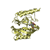
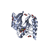
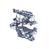
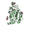
| Title | Structure of the Mutant D206N of Phycoerythrobilin Synthase PebS from the Cyanophage P-SSM2 in complex with bound substrate Biliverdin IXA | ||||||
 Components Components | PHYCOERYTHROBILIN SYNTHASE | ||||||
 Keywords Keywords | OXIDOREDUCTASE | ||||||
| Function / homology |  Function and homology information Function and homology informationphycoerythrobilin synthase / phytochromobilin biosynthetic process / oxidoreductase activity, acting on the CH-CH group of donors, iron-sulfur protein as acceptor / cobalt ion binding Similarity search - Function | ||||||
| Biological species |  PROCHLOROCOCCUS PHAGE P-SSM2 (virus) PROCHLOROCOCCUS PHAGE P-SSM2 (virus) | ||||||
| Method |  X-RAY DIFFRACTION / SYNCHROTRON / MOLECULAR REPLACEMENT / Resolution: 1.85 Å X-RAY DIFFRACTION / SYNCHROTRON / MOLECULAR REPLACEMENT / Resolution: 1.85 Å | ||||||
 Authors Authors | Busch, A.W.U. / Frankenberg-Dinkel, N. / Hofmann, E. | ||||||
 Citation Citation | Journal: Biochem.J. / Year: 2011 Title: Radical Mechanism of Cyanophage Phycoerythrobilin Synthase (Pebs). Authors: Busch, A.W.U. / Reijerse, E.J. / Lubitz, W. / Hofmann, E. / Frankenberg-Dinkel, N. | ||||||
| History |
|
- Structure visualization
| Structure viewer | Molecule: MolmilJmol/JSmol |
|---|
- Downloads & links
-Download
| PDBx/mmCIF format | 2x9j.cif.gz | 112.7 KB | Display | PDBx/mmCIF format |
|---|---|---|---|---|
| PDB format | pdb2x9j.ent.gz | 86.3 KB | Display | PDB format |
| PDBx/mmJSON format | 2x9j.json.gz | Tree view | PDBx/mmJSON format | |
| Others |  Other downloads Other downloads |
-Validation report
| Arichive directory | https://data.pdbj.org/pub/pdb/validation_reports/x9/2x9jftp://data.pdbj.org/pub/pdb/validation_reports/x9/2x9j | HTTPS FTP |
|---|
-Related structure data
| Related structure data |  2x9iC  2vgrS C: citing same article ( S: Starting model for refinement |
|---|---|
| Similar structure data |


-Links
 PDBj
PDBj- Assembly
| Deposited unit | 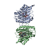
| ||||||||||||||||||||||||||||||||||||||||||||||
|---|---|---|---|---|---|---|---|---|---|---|---|---|---|---|---|---|---|---|---|---|---|---|---|---|---|---|---|---|---|---|---|---|---|---|---|---|---|---|---|---|---|---|---|---|---|---|---|
| 1 | 
| ||||||||||||||||||||||||||||||||||||||||||||||
| 2 | 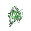
| ||||||||||||||||||||||||||||||||||||||||||||||
| Unit cell |
| ||||||||||||||||||||||||||||||||||||||||||||||
| Noncrystallographic symmetry (NCS) | NCS domain:
NCS domain segments:
NCS ensembles :
NCS oper: (Code: given Matrix: (-0.990974, -0.134053, -0.000499), Vector: |
-Components
| #1: Protein | Mass: 27291.855 Da / Num. of mol.: 2 / Mutation: YES Source method: isolated from a genetically manipulated source Source: (gene. exp.) PROCHLOROCOCCUS PHAGE P-SSM2 (virus) / Description: CYANOPHAGE / Plasmid: PGEX-6P-3 / Production host:  #2: Chemical |   Mass: 582.646 Da / Num. of mol.: 2 / Source method: obtained synthetically / Formula: C33H34N4O6 Mass: 582.646 Da / Num. of mol.: 2 / Source method: obtained synthetically / Formula: C33H34N4O6#3: Water | ChemComp-HOH / |  Mass: 18.015 Da / Num. of mol.: 429 / Source method: isolated from a natural source / Formula: H2O Mass: 18.015 Da / Num. of mol.: 429 / Source method: isolated from a natural source / Formula: H2OCompound details | ENGINEERED | |
|---|
-Experimental details
-Experiment
| Experiment | Method: X-RAY DIFFRACTION / Number of used crystals: 1 |
|---|
- Sample preparation
| Crystal | Density Matthews: 2.41 Å3/Da / Density % sol: 49 % / Description: NONE |
|---|---|
| Crystal grow | Details: 170MM (NH4)2SO4, 20%PEG3350 |
-Data collection
| Diffraction | Mean temperature: 100 K |
|---|---|
| Diffraction source | Source: SYNCHROTRON / Site: ESRF  / Beamline: ID23-2 / Wavelength: 0.8726 / Beamline: ID23-2 / Wavelength: 0.8726 |
| Detector | Type: MARRESEARCH / Detector: CCD / Date: Oct 2, 2008 / Details: MIRROR |
| Radiation | Monochromator: SILICON 111 CRYSTAL / Protocol: SINGLE WAVELENGTH / Monochromatic (M) / Laue (L): M / Scattering type: x-ray |
| Radiation wavelength | Wavelength: 0.8726 Å / Relative weight: 1 |
| Reflection | Resolution: 1.85→49 Å / Num. obs: 44404 / % possible obs: 99.8 % / Observed criterion σ(I): 0 / Redundancy: 5.88 % / Biso Wilson estimate: 26.2 Å2 / Rmerge(I) obs: 0.08 / Net I/σ(I): 16.2 |
| Reflection shell | Resolution: 1.85→1.9 Å / Redundancy: 5.93 % / Rmerge(I) obs: 0.45 / Mean I/σ(I) obs: 4.26 / % possible all: 99.7 |
- Processing
| Software |
| |||||||||||||||||||||||||||||||||||||||||||||||||||||||||||||||||||||||||||||||||||||||||||||||||||||||||||||||||||||||
|---|---|---|---|---|---|---|---|---|---|---|---|---|---|---|---|---|---|---|---|---|---|---|---|---|---|---|---|---|---|---|---|---|---|---|---|---|---|---|---|---|---|---|---|---|---|---|---|---|---|---|---|---|---|---|---|---|---|---|---|---|---|---|---|---|---|---|---|---|---|---|---|---|---|---|---|---|---|---|---|---|---|---|---|---|---|---|---|---|---|---|---|---|---|---|---|---|---|---|---|---|---|---|---|---|---|---|---|---|---|---|---|---|---|---|---|---|---|---|---|---|
| Refinement | Method to determine structure: MOLECULAR REPLACEMENT Starting model: PDB ENTRY 2VGR Resolution: 1.85→39.687 Å / SU ML: 0.19 / σ(F): 2 / Phase error: 20.47 / Stereochemistry target values: ML
| |||||||||||||||||||||||||||||||||||||||||||||||||||||||||||||||||||||||||||||||||||||||||||||||||||||||||||||||||||||||
| Solvent computation | Shrinkage radii: 0.9 Å / VDW probe radii: 1.11 Å / Solvent model: FLAT BULK SOLVENT MODEL / Bsol: 40.26 Å2 / ksol: 0.335 e/Å3 | |||||||||||||||||||||||||||||||||||||||||||||||||||||||||||||||||||||||||||||||||||||||||||||||||||||||||||||||||||||||
| Displacement parameters |
| |||||||||||||||||||||||||||||||||||||||||||||||||||||||||||||||||||||||||||||||||||||||||||||||||||||||||||||||||||||||
| Refinement step | Cycle: LAST / Resolution: 1.85→39.687 Å
| |||||||||||||||||||||||||||||||||||||||||||||||||||||||||||||||||||||||||||||||||||||||||||||||||||||||||||||||||||||||
| Refine LS restraints |
| |||||||||||||||||||||||||||||||||||||||||||||||||||||||||||||||||||||||||||||||||||||||||||||||||||||||||||||||||||||||
| Refine LS restraints NCS |
| |||||||||||||||||||||||||||||||||||||||||||||||||||||||||||||||||||||||||||||||||||||||||||||||||||||||||||||||||||||||
| LS refinement shell |
|
