[English] 日本語
Yorodumi
- PDB-2fmd: Structural basis of carbohydrate recognition by Bowringia milbrae... -

+
Open data


ID or keywords:

Loading...

-
Basic information

Entry
Database: PDB / ID: 2fmd
TitleStructural basis of carbohydrate recognition by Bowringia milbraedii seed agglutinin
ComponentsLectin
KeywordsSUGAR BINDING PROTEIN / LEGUME LECTIN / BETA SANDWICH / PROTEIN-CARBOHYDRATE COMPLEX / lectin
Function / homology
Function and homology information


carbohydrate binding / metal ion binding
Similarity search - Function
Legume lectin / Legume lectins beta-chain signature. / Legume lectin domain / Legume lectin, beta chain, Mn/Ca-binding site / Legume lectin domain / Jelly Rolls - #200 / Concanavalin A-like lectin/glucanase domain superfamily / Jelly Rolls / Sandwich / Mainly Beta
Similarity search - Domain/homology
2alpha-alpha-mannobiose / : / Lectin
Similarity search - Component
Biological speciesBowringia mildbraedii (plant)
MethodX-RAY DIFFRACTION / SYNCHROTRON / MOLECULAR REPLACEMENT / Resolution: 1.9 Å
AuthorsButs, L. / Garcia-Pino, A. / Wyns, L. / Loris, R.
Citation
Journal: Glycobiology / Year: 2006
Title: Structural basis of carbohydrate recognition by a Man(alpha1-2)Man-specific lectin from Bowringia milbraedii.
Authors: Buts, L. / Garcia-Pino, A. / Wyns, L. / Loris, R.
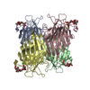
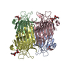
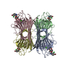
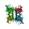
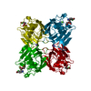
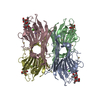
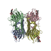
#1: Journal: Acta Crystallogr.,Sect.F / Year: 2005
Title: Crystallization and preliminary X-ray analysis of the Man(alpha 1-2)Man-specific lectin from Bowringia mildbraedii in complex with its carbohydrate ligand
History
DepositionJan 9, 2006Deposition site: RCSB / Processing site: RCSB
Revision 1.0Aug 22, 2006Provider: repository / Type: Initial release
Revision 1.1May 1, 2008Group: Version format compliance
Revision 1.2Jul 13, 2011Group: Version format compliance
Revision 2.0Jul 29, 2020Group: Atomic model / Data collection ...Atomic model / Data collection / Derived calculations / Structure summary
Category: atom_site / chem_comp ...atom_site / chem_comp / diffrn_source / entity / entity_name_com / pdbx_branch_scheme / pdbx_chem_comp_identifier / pdbx_entity_branch / pdbx_entity_branch_descriptor / pdbx_entity_branch_link / pdbx_entity_branch_list / pdbx_entity_nonpoly / pdbx_molecule_features / pdbx_nonpoly_scheme / pdbx_struct_assembly_gen / pdbx_struct_conn_angle / struct_asym / struct_conn / struct_site / struct_site_gen
Item: _atom_site.B_iso_or_equiv / _atom_site.Cartn_x ..._atom_site.B_iso_or_equiv / _atom_site.Cartn_x / _atom_site.Cartn_y / _atom_site.Cartn_z / _atom_site.auth_asym_id / _atom_site.auth_atom_id / _atom_site.auth_seq_id / _atom_site.label_asym_id / _atom_site.label_atom_id / _atom_site.type_symbol / _chem_comp.name / _chem_comp.type / _diffrn_source.pdbx_synchrotron_site / _entity.formula_weight / _entity.pdbx_description / _entity.pdbx_number_of_molecules / _entity.type / _pdbx_struct_assembly_gen.asym_id_list / _pdbx_struct_conn_angle.ptnr1_auth_comp_id / _pdbx_struct_conn_angle.ptnr1_auth_seq_id / _pdbx_struct_conn_angle.ptnr1_label_asym_id / _pdbx_struct_conn_angle.ptnr1_label_atom_id / _pdbx_struct_conn_angle.ptnr1_label_comp_id / _pdbx_struct_conn_angle.ptnr1_label_seq_id / _pdbx_struct_conn_angle.ptnr2_auth_comp_id / _pdbx_struct_conn_angle.ptnr2_auth_seq_id / _pdbx_struct_conn_angle.ptnr2_label_asym_id / _pdbx_struct_conn_angle.ptnr2_label_atom_id / _pdbx_struct_conn_angle.ptnr2_label_comp_id / _pdbx_struct_conn_angle.ptnr3_auth_comp_id / _pdbx_struct_conn_angle.ptnr3_auth_seq_id / _pdbx_struct_conn_angle.ptnr3_label_asym_id / _pdbx_struct_conn_angle.ptnr3_label_atom_id / _pdbx_struct_conn_angle.ptnr3_label_comp_id / _pdbx_struct_conn_angle.ptnr3_label_seq_id / _pdbx_struct_conn_angle.value / _struct_conn.pdbx_dist_value / _struct_conn.pdbx_leaving_atom_flag / _struct_conn.ptnr1_auth_asym_id / _struct_conn.ptnr1_auth_comp_id / _struct_conn.ptnr1_auth_seq_id / _struct_conn.ptnr1_label_asym_id / _struct_conn.ptnr1_label_atom_id / _struct_conn.ptnr1_label_comp_id / _struct_conn.ptnr1_label_seq_id / _struct_conn.ptnr2_auth_asym_id / _struct_conn.ptnr2_auth_comp_id / _struct_conn.ptnr2_auth_seq_id / _struct_conn.ptnr2_label_asym_id / _struct_conn.ptnr2_label_atom_id / _struct_conn.ptnr2_label_comp_id / _struct_conn.ptnr2_label_seq_id
Description: Carbohydrate remediation / Provider: repository / Type: Remediation
Revision 2.1Aug 30, 2023Group: Data collection / Database references ...Data collection / Database references / Refinement description / Structure summary
Category: chem_comp / chem_comp_atom ...chem_comp / chem_comp_atom / chem_comp_bond / database_2 / pdbx_initial_refinement_model
Item: _chem_comp.pdbx_synonyms / _database_2.pdbx_DOI / _database_2.pdbx_database_accession
Remark 999SEQUENCE ALL THESE CONFLICS ARE DUE TO CLEAR DISCREPANCIES BETWEEN THE DATABASE SEQUENCE AND THE ...SEQUENCE ALL THESE CONFLICS ARE DUE TO CLEAR DISCREPANCIES BETWEEN THE DATABASE SEQUENCE AND THE OBSERVED ELECTRON DENSITY. WE THEREFORE BELIEVE THAT THE CORRESPONDING RESIDUES IN THE PUBLISHED SEQUENCE ARE INCORRECT. THIS IS PARTICULARLY IMPORTANT FOR ASP86, WHICH IS A CONSERVED RESIDUE AND ABSOLUTELY ESSENTIAL FOR THE LECTIN'S FUNCTION. IN THE CASE OF RESIDUES 204 AND 206 AUTHORS WERE UNABLE TO DISTINGUISH ASP AND ASN FROM THE DENSITY, HENCE THE ASX RESIDUES.

-
Structure visualization

Structure viewerMolecule:
MolmilJmol/JSmol

Downloads & links

-
Assembly

Deposited unit
A: Lectin
hetero molecules


Theoretical massNumber of molelcules
Total (without water)26,0204
Polymers25,5831
Non-polymers4373
Water3,837213
1
A: Lectin
hetero molecules

A: Lectin
hetero molecules

A: Lectin
hetero molecules

A: Lectin
hetero molecules


Theoretical massNumber of molelcules
Total (without water)104,08116
Polymers102,3324
Non-polymers1,74912
Water724
TypeNameSymmetry operationNumber
identity operation1_555x,y,z1
crystal symmetry operation2_555-x,-y,z1
crystal symmetry operation3_555-x,y,-z1
crystal symmetry operation4_555x,-y,-z1
Unit cell
Length a, b, c (Å)66.603, 86.351, 91.760
Angle α, β, γ (deg.)90.00, 90.00, 90.00
Int Tables number23
Space group name H-MI222
DetailsTetramer assembled using the crystallographic symmetry operators (-X,-Y,Z), (X,-Y,-Z) and (-X,Y,-Z)

-
Components

#1: Protein Lectin / Agglutinin / BMA


Mass: 25582.971 Da / Num. of mol.: 1 / Source method: isolated from a natural source / Source: (natural) Bowringia mildbraedii (plant) / Tissue: seed / References: UniProt: P42088
#2: Polysaccharide alpha-D-mannopyranose-(1-2)-alpha-D-mannopyranose / 2alpha-alpha-mannobiose


Type: oligosaccharide, Oligosaccharide / Class: Metabolism / Mass: 342.297 Da / Num. of mol.: 1
Source method: isolated from a genetically manipulated source
Details: oligosaccharide / References: 2alpha-alpha-mannobiose
DescriptorTypeProgram
DManpa1-2DManpa1-ROHGlycam Condensed SequenceGMML 1.0
WURCS=2.0/1,2,1/[a1122h-1a_1-5]/1-1/a2-b1WURCSPDB2Glycan 1.1.0
[][a-D-Manp]{[(2+1)][a-D-Manp]{}}LINUCSPDB-CARE
#3: Chemical ChemComp-CA / CALCIUM ION


Mass: 40.078 Da / Num. of mol.: 1 / Source method: obtained synthetically / Formula: Ca
#4: Chemical ChemComp-MN / MANGANESE (II) ION


Mass: 54.938 Da / Num. of mol.: 1 / Source method: obtained synthetically / Formula: Mn
#5: Water ChemComp-HOH / water


Mass: 18.015 Da / Num. of mol.: 213 / Source method: isolated from a natural source / Formula: H2O

-
Experimental details

-
Experiment

ExperimentMethod: X-RAY DIFFRACTION / Number of used crystals: 1

-
Sample preparation

CrystalDensity Matthews: 2.55 Å3/Da / Density % sol: 51.84 %
Crystal growTemperature: 277 K / Method: vapor diffusion, hanging drop / pH: 6.5
Details: 15% PEG 20000, 0.1M MES pH 6.5, VAPOR DIFFUSION, HANGING DROP, temperature 277K

-
Data collection

DiffractionMean temperature: 100 K
Diffraction sourceSource: SYNCHROTRON / Site: EMBL/DESY, HAMBURG / Beamline: X11 / Wavelength: 0.8123 Å
DetectorType: MARRESEARCH / Detector: CCD / Date: Jun 8, 2005 / Details: mirrors
RadiationProtocol: SINGLE WAVELENGTH / Monochromatic (M) / Laue (L): M / Scattering type: x-ray
Radiation wavelengthWavelength: 0.8123 Å / Relative weight: 1
ReflectionResolution: 1.9→15 Å / Num. all: 18493 / Num. obs: 18493 / % possible obs: 87.3 % / Observed criterion σ(F): 0 / Observed criterion σ(I): 0 / Redundancy: 5.4 % / Rmerge(I) obs: 0.095

-
Processing

Software
NameVersionClassification
DENZOdata reduction
SCALEPACKdata scaling
PHASERphasing
CNS1.1refinement
RefinementMethod to determine structure: MOLECULAR REPLACEMENT
Starting model: PDB ENTRY 1Q8S, CHAIN A
Resolution: 1.9→15 Å / Isotropic thermal model: Isotropic / Cross valid method: THROUGHOUT / σ(F): 0 / σ(I): 0 / Stereochemistry target values: Engh & Huber
RfactorNum. reflection% reflectionSelection details
Rfree0.228 1450 -RANDOM
Rwork0.183 ---
all0.186 18493 --
obs0.186 17043 80.4 %-
Refinement stepCycle: LAST / Resolution: 1.9→15 Å
ProteinNucleic acidLigandSolventTotal
Num. atoms1736 0 25 213 1974
Refine LS restraints
Refine-IDTypeDev ideal
X-RAY DIFFRACTIONc_angle_d1.674
X-RAY DIFFRACTIONc_bond_d0.008

+
About Yorodumi

-
News

-
Feb 9, 2022. New format data for meta-information of EMDB entries

New format data for meta-information of EMDB entries

  • Version 3 of the EMDB header file is now the official format.
  • The previous official version 1.9 will be removed from the archive.

Related info.:EMDB header

External links:wwPDB to switch to version 3 of the EMDB data model

-
Aug 12, 2020. Covid-19 info

Covid-19 info

URL: https://pdbjlvh1.pdbj.org/emnavi/covid19.php

New page: Covid-19 featured information page in EM Navigator.

Related info.:Covid-19 info / Mar 5, 2020. Novel coronavirus structure data

+
Mar 5, 2020. Novel coronavirus structure data

Novel coronavirus structure data

Related info.:Yorodumi Speices / Aug 12, 2020. Covid-19 info

External links:COVID-19 featured content - PDBj / Molecule of the Month (242):Coronavirus Proteases

+
Jan 31, 2019. EMDB accession codes are about to change! (news from PDBe EMDB page)

EMDB accession codes are about to change! (news from PDBe EMDB page)

  • The allocation of 4 digits for EMDB accession codes will soon come to an end. Whilst these codes will remain in use, new EMDB accession codes will include an additional digit and will expand incrementally as the available range of codes is exhausted. The current 4-digit format prefixed with “EMD-” (i.e. EMD-XXXX) will advance to a 5-digit format (i.e. EMD-XXXXX), and so on. It is currently estimated that the 4-digit codes will be depleted around Spring 2019, at which point the 5-digit format will come into force.
  • The EM Navigator/Yorodumi systems omit the EMD- prefix.

Related info.:Q: What is EMD? / ID/Accession-code notation in Yorodumi/EM Navigator

External links:EMDB Accession Codes are Changing Soon! / Contact to PDBj

+
Jul 12, 2017. Major update of PDB

Major update of PDB

  • wwPDB released updated PDB data conforming to the new PDBx/mmCIF dictionary.
  • This is a major update changing the version number from 4 to 5, and with Remediation, in which all the entries are updated.
  • In this update, many items about electron microscopy experimental information are reorganized (e.g. em_software).
  • Now, EM Navigator and Yorodumi are based on the updated data.

External links:wwPDB Remediation / Enriched Model Files Conforming to OneDep Data Standards Now Available in the PDB FTP Archive

-
Yorodumi

Thousand views of thousand structures

  • Yorodumi is a browser for structure data from EMDB, PDB, SASBDB, etc.
  • This page is also the successor to EM Navigator detail page, and also detail information page/front-end page for Omokage search.
  • The word "yorodu" (or yorozu) is an old Japanese word meaning "ten thousand". "mi" (miru) is to see.

Related info.:EMDB / PDB / SASBDB / Comparison of 3 databanks / Yorodumi Search / Aug 31, 2016. New EM Navigator & Yorodumi / Yorodumi Papers / Jmol/JSmol / Function and homology information / Changes in new EM Navigator and Yorodumi

Read more