 Movie
Movie Controller

[English] 日本語
 Yorodumi
- PDB-2wtx: Insight into the mechanism of enzymatic glycosyltransfer with ret... -
+ Open data

- Basic information
| Entry | Database: PDB / ID: 2wtx | ||||||
|---|---|---|---|---|---|---|---|
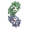
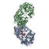
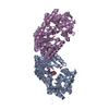
| Title | Insight into the mechanism of enzymatic glycosyltransfer with retention through the synthesis and analysis of bisubstrate glycomimetics of trehalose-6-phosphate synthase | ||||||
 Components Components | ALPHA, ALPHA-TREHALOSE-PHOSPHATE SYNTHASE [UDP-FORMING] | ||||||
 Keywords Keywords | TRANSFERASE / TREHALOSE SYNTHASE / GLYCOSYLTRANSFERASE / POTASSIUM / RETENTION / STRESS RESPONSE | ||||||
| Function / homology |  Function and homology information Function and homology informationalpha,alpha-trehalose-phosphate synthase (UDP-forming) / alpha,alpha-trehalose-phosphate synthase (UDP-forming) activity / trehalose biosynthetic process / response to osmotic stress / cellular response to cold / response to stress / DNA damage response Similarity search - Function | ||||||
| Biological species |  | ||||||
| Method |  X-RAY DIFFRACTION / SYNCHROTRON / MOLECULAR REPLACEMENT / Resolution: 2.2 Å X-RAY DIFFRACTION / SYNCHROTRON / MOLECULAR REPLACEMENT / Resolution: 2.2 Å | ||||||
 Authors Authors | Errey, J.C. / Lee, S.S. / Gibson, R.P. / Martinez-Fleites, C. / Barry, C.S. / Jung, P.M.J. / OSullivan, A. / Davis, B.G. / Davies, G.J. | ||||||
 Citation Citation | Journal: Angew.Chem.Int.Ed.Engl. / Year: 2010 Title: Mechanistic Insight Into Enzymatic Glycosyl Transfer with Retention of Configuration Through Analysis of Glycomimetic Inhibitors. Authors: Errey, J.C. / Lee, S.S. / Gibson, R.P. / Martinez Fleites, C. / Barry, C.S. / Jung, P.M.J. / O'Sullivan, A.C. / Davis, B.G. / Davies, G.J. | ||||||
| History |
|
- Structure visualization
| Structure viewer | Molecule: MolmilJmol/JSmol |
|---|
- Downloads & links
-Download
| PDBx/mmCIF format | 2wtx.cif.gz | 379.4 KB | Display | PDBx/mmCIF format |
|---|---|---|---|---|
| PDB format | pdb2wtx.ent.gz | 310.5 KB | Display | PDB format |
| PDBx/mmJSON format | 2wtx.json.gz | Tree view | PDBx/mmJSON format | |
| Others |  Other downloads Other downloads |
-Validation report
| Arichive directory | https://data.pdbj.org/pub/pdb/validation_reports/wt/2wtxftp://data.pdbj.org/pub/pdb/validation_reports/wt/2wtx | HTTPS FTP |
|---|
-Related structure data
| Related structure data | 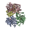 1gz5S S: Starting model for refinement |
|---|---|
| Similar structure data |


-Links
 PDBj
PDBj


- Assembly
| Deposited unit | 
| ||||||||
|---|---|---|---|---|---|---|---|---|---|
| 1 | 
| ||||||||
| 2 | 
| ||||||||
| Unit cell |
|
-Components
| #1: Protein | Mass: 53675.973 Da / Num. of mol.: 4 Source method: isolated from a genetically manipulated source Source: (gene. exp.) References: UniProt: P31677, alpha,alpha-trehalose-phosphate synthase (UDP-forming) #2: Chemical | ChemComp-EDO /   Mass: 62.068 Da / Num. of mol.: 5 / Source method: obtained synthetically / Formula: C2H6O2 Mass: 62.068 Da / Num. of mol.: 5 / Source method: obtained synthetically / Formula: C2H6O2#3: Chemical | ChemComp-UDP /   Type: RNA linking / Mass: 404.161 Da / Num. of mol.: 4 / Source method: obtained synthetically / Formula: C9H14N2O12P2 / Comment: UDP*YM Type: RNA linking / Mass: 404.161 Da / Num. of mol.: 4 / Source method: obtained synthetically / Formula: C9H14N2O12P2 / Comment: UDP*YM#4: Chemical | ChemComp-VDO / [(   Mass: 415.330 Da / Num. of mol.: 4 / Source method: obtained synthetically / Formula: C14H26NO11P Mass: 415.330 Da / Num. of mol.: 4 / Source method: obtained synthetically / Formula: C14H26NO11P#5: Water | ChemComp-HOH / |  Mass: 18.015 Da / Num. of mol.: 585 / Source method: isolated from a natural source / Formula: H2O Mass: 18.015 Da / Num. of mol.: 585 / Source method: isolated from a natural source / Formula: H2O |
|---|
-Experimental details
-Experiment
| Experiment | Method: X-RAY DIFFRACTION / Number of used crystals: 1 |
|---|
- Sample preparation
| Crystal | Density Matthews: 2.55 Å3/Da / Density % sol: 51 % / Description: NONE |
|---|
-Data collection
| Diffraction | Mean temperature: 100 K |
|---|---|
| Diffraction source | Source: SYNCHROTRON / Site: ESRF  / Beamline: ID14-1 / Wavelength: 0.9791 / Beamline: ID14-1 / Wavelength: 0.9791 |
| Detector | Type: ADSC CCD / Detector: CCD |
| Radiation | Protocol: SINGLE WAVELENGTH / Monochromatic (M) / Laue (L): M / Scattering type: x-ray |
| Radiation wavelength | Wavelength: 0.9791 Å / Relative weight: 1 |
| Reflection | Resolution: 2.2→99 Å / Num. obs: 111567 / % possible obs: 99.9 % / Observed criterion σ(I): 2 / Redundancy: 5.2 % / Biso Wilson estimate: 30 Å2 / Rmerge(I) obs: 0.1 / Net I/σ(I): 14 |
| Reflection shell | Resolution: 2.2→2.3 Å / Redundancy: 3.7 % / Rmerge(I) obs: 0.34 / Mean I/σ(I) obs: 5 / % possible all: 99.4 |
- Processing
| Software |
| ||||||||||||||||||||||||||||||||||||||||||||||||||||||||||||||||||||||||||||||||||||||||||||||||||||||||||||||||||||||||||||||||||||||||||||||||||||||||||||||||||||||||||||||||||||||
|---|---|---|---|---|---|---|---|---|---|---|---|---|---|---|---|---|---|---|---|---|---|---|---|---|---|---|---|---|---|---|---|---|---|---|---|---|---|---|---|---|---|---|---|---|---|---|---|---|---|---|---|---|---|---|---|---|---|---|---|---|---|---|---|---|---|---|---|---|---|---|---|---|---|---|---|---|---|---|---|---|---|---|---|---|---|---|---|---|---|---|---|---|---|---|---|---|---|---|---|---|---|---|---|---|---|---|---|---|---|---|---|---|---|---|---|---|---|---|---|---|---|---|---|---|---|---|---|---|---|---|---|---|---|---|---|---|---|---|---|---|---|---|---|---|---|---|---|---|---|---|---|---|---|---|---|---|---|---|---|---|---|---|---|---|---|---|---|---|---|---|---|---|---|---|---|---|---|---|---|---|---|---|---|
| Refinement | Method to determine structure: MOLECULAR REPLACEMENT Starting model: PDB ENTRY 1GZ5 Resolution: 2.2→98.91 Å / Cor.coef. Fo:Fc: 0.937 / Cor.coef. Fo:Fc free: 0.911 / SU B: 5.365 / SU ML: 0.139 / Cross valid method: THROUGHOUT / ESU R: 0.257 / ESU R Free: 0.2 / Stereochemistry target values: MAXIMUM LIKELIHOOD Details: HYDROGENS HAVE BEEN ADDED IN THE RIDING POSITIONS. U VALUES REFINED INDIVIDUALLY.
| ||||||||||||||||||||||||||||||||||||||||||||||||||||||||||||||||||||||||||||||||||||||||||||||||||||||||||||||||||||||||||||||||||||||||||||||||||||||||||||||||||||||||||||||||||||||
| Solvent computation | Ion probe radii: 0.8 Å / Shrinkage radii: 0.8 Å / VDW probe radii: 1.2 Å / Solvent model: MASK | ||||||||||||||||||||||||||||||||||||||||||||||||||||||||||||||||||||||||||||||||||||||||||||||||||||||||||||||||||||||||||||||||||||||||||||||||||||||||||||||||||||||||||||||||||||||
| Displacement parameters | Biso mean: 29.981 Å2
| ||||||||||||||||||||||||||||||||||||||||||||||||||||||||||||||||||||||||||||||||||||||||||||||||||||||||||||||||||||||||||||||||||||||||||||||||||||||||||||||||||||||||||||||||||||||
| Refinement step | Cycle: LAST / Resolution: 2.2→98.91 Å
| ||||||||||||||||||||||||||||||||||||||||||||||||||||||||||||||||||||||||||||||||||||||||||||||||||||||||||||||||||||||||||||||||||||||||||||||||||||||||||||||||||||||||||||||||||||||
| Refine LS restraints |
|